
You’ll find 3D bioprinted scaffolds for tendon repair are revolutionary tissue engineering constructs that combine living cells, bioactive materials, and precisely designed structures to restore damaged tendons. These scaffolds use advanced bioprinting techniques like extrusion-based and inkjet printing to create patient-specific treatments that replicate natural tendon architecture. They address the high failure rates of traditional surgical methods by providing better mechanical strength and biological functionality. Discover how these innovations are transforming tendon recovery and offering new hope for patients.
Understanding Tendon Structure and Function in the Musculoskeletal System
Although tendons make up only a small fraction of your body’s total mass, they’re essential connective tissues that transmit forces from muscles to bones while helping maintain your posture throughout the musculoskeletal system.
Your tendon structure consists primarily of collagen type I, which forms hierarchically arranged fibrils within a hydrated extracellular matrix. Specialized cells called tenocytes continuously synthesize and maintain this matrix, ensuring your tendons retain their structural integrity.
Tenocytes continuously synthesize collagen type I fibrils within your tendon’s hydrated matrix to maintain essential structural integrity.
The mechanical properties of your tendons exhibit viscoelastic behavior, meaning they respond differently to varying strain rates.
Unfortunately, tendon injuries are common and often challenging to heal. Chronic conditions like tendinopathy persist partly because we’re still developing our understanding of tendon biology.
Current Challenges in Traditional Tendon Repair Methods
When tendon injuries occur, traditional repair methods present significant limitations that can compromise your recovery and long-term outcomes.
You’ll face alarmingly high retear rates, with surgical treatments showing up to 25% failure in patients over 70. Non-surgical approaches often can’t restore adequate structural integrity for severe tears or complete ruptures.
When you require grafts for extensively damaged tendons, these techniques frequently fail to restore full functionality and may cause complications.
Chronic tendon injuries particularly challenge traditional methods, as current approaches don’t effectively address complex tendon-bone healing processes.
Your healing becomes compromised due to limited understanding of tendon biology and ineffective regeneration mechanisms.
These shortcomings highlight why researchers are developing innovative solutions like 3D bioprinted scaffolds for tendon repair, offering hope for improved outcomes.
Fundamentals of 3D Bioprinting Technology for Tissue Engineering
When you’re exploring 3D bioprinting for tendon repair, you’ll encounter several distinct printing methods including extrusion-based, inkjet, laser-assisted bioprinting, and stereolithography, each offering unique advantages in resolution and cell compatibility.
You can leverage digital design capabilities to create precise, patient-specific scaffolds that match the exact geometry and mechanical requirements of damaged tendons. This technology allows you to control scaffold architecture at the microscale while incorporating living cells and biomaterials in a layer-by-layer construction process.
Bioprinting Methods Overview
Since traditional tissue engineering approaches often fall short of replicating tendon’s complex architecture, 3D bioprinting has emerged as a revolutionary technology that enables precise spatial control over cell placement and scaffold geometry.
You’ll find three primary bioprinting techniques available for tendon tissue engineering.
Extrusion printing allows you to create high-viscosity scaffolds by squeezing bioink through nozzles, though it may compromise cell viability.
Inkjet printing offers superior resolution through droplet deposition but limits cell density in your constructs.
Laser-assisted bioprinting provides the best solution, avoiding blockage issues while maintaining higher cell viability.
Each method enables you to incorporate living cells and bioactive molecules into scaffolds, promoting the cell adhesion and proliferation essential for successful tendon regeneration.
Digital Design Advantages
The real power of 3D bioprinting lies in its digital foundation, which transforms tendon repair from a one-size-fits-all approach to precision medicine. You can customize scaffolds with computer-aided design modeling, controlling compositions and internal structures to mimic natural tendon architecture. Digital design enables integration of living cells and biomaterials into complex geometric structures that enhance tendon regeneration.
| Digital Design Feature | Benefit | Application |
|---|---|---|
| Controllable Compositions | Precise material placement | Hierarchical collagen arrangement |
| Cell Encapsulation | Enhanced biocompatibility | Growth factor delivery |
| Multilayered Architecture | Improved mechanical properties | Biomimetic scaffolds |
| Rapid Prototyping | Faster development cycles | Customized patient solutions |
You’ll achieve reproducible, biomimetic scaffolds that closely replicate natural tendon properties through this precision-driven approach.
Inkjet Bioprinting Techniques for Tendon Scaffold Creation
Although traditional manufacturing methods struggle with creating intricate tendon architectures, inkjet bioprinting offers a revolutionary approach that precisely deposits bioink droplets to form complex three-dimensional scaffolds.
You’ll benefit from this technique’s exceptional resolution of less than 30 µm, enabling precise control over scaffold geometry that mimics natural tendon structures essential for effective regeneration.
However, you’ll need to address challenges like low cell density from limited droplet volumes, which can impact cell viability and mechanical strength.
The technique’s ability to layer multiple bioinks allows you to incorporate different cell types and bioactive molecules into your tendon scaffolds.
Continuous innovations focus on optimizing bioink viscosity and reducing shear stress to maintain cell viability throughout the printing process.
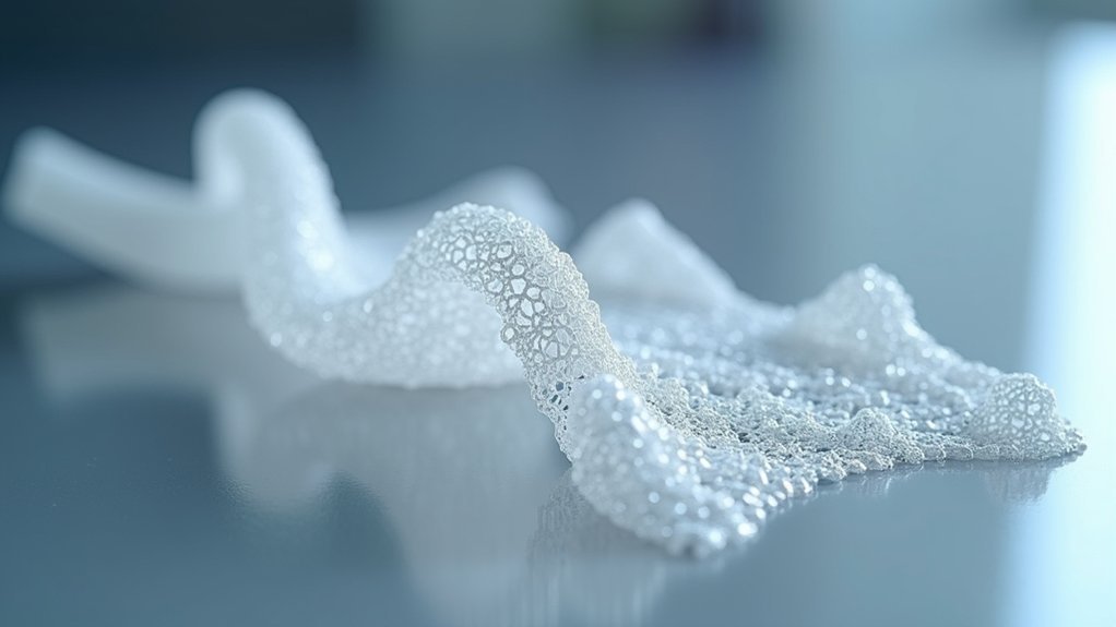
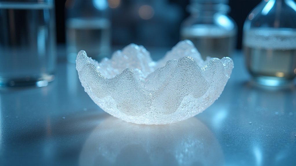
Extrusion-Based Bioprinting Methods and Applications
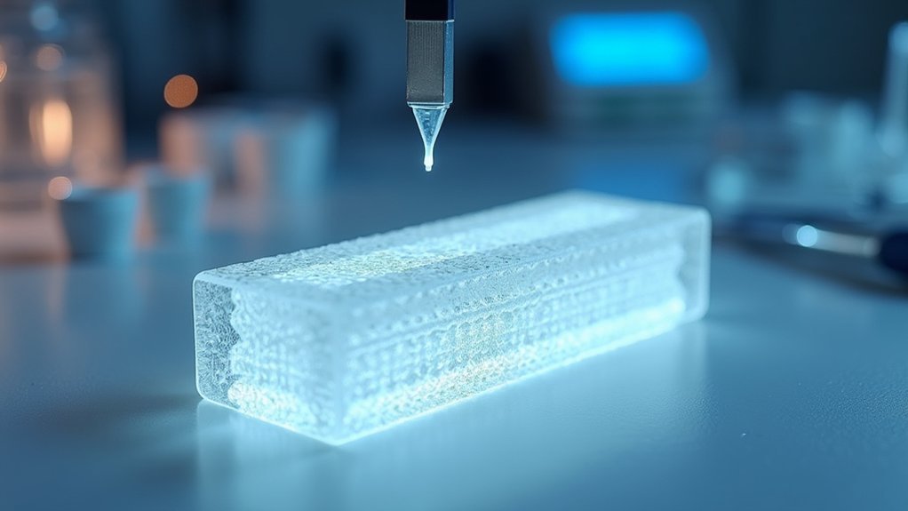
When you’re working with extrusion-based bioprinting, you’ll control the bioink deposition process by adjusting nozzle pressure and flow rates to create precise scaffold geometries.
You’ll face significant cell viability challenges since the high-pressure extrusion forces can damage living cells during the printing process.
You’ll need to master material distribution control techniques to guarantee uniform cell seeding and prevent aggregation throughout your multi-layered tendon scaffolds.
Bioink Deposition Process
Extrusion-based bioprinting fundamentally operates by depositing bioink through a nozzle under controlled pressure, creating layer-by-layer tissue scaffolds that can accommodate high-viscosity formulations essential for structural integrity.
During the bioink deposition process, you’ll need precise control over pressure (1.5–2.0 bar), speed, and interlayer distance to guarantee consistent extrusion and proper scaffold dimensions.
This extrusion-based bioprinting method enables you to create complex geometries and multi-layered structures that mimic natural tendon organization, enhancing mechanical properties while supporting cell attachment.
You can incorporate sacrificial materials like Pluronic F127 to provide structural support during printing, which you’ll later remove to create porous architectures.
However, you must balance high-viscosity benefits for scaffold structure against potential impacts on cell viability and printing resolution.
Cell Viability Challenges
While bioink deposition offers structural advantages, you’ll encounter significant cell viability challenges that can compromise your scaffold’s biological functionality. The extrusion process generates high shear stress that damages sensitive cells, reducing their survival rates and functionality. High-viscosity bioinks worsen these issues by creating non-uniform cell distribution throughout your printed scaffolds.
| Challenge | Solution |
|---|---|
| High shear stress during extrusion | Optimize nozzle design and printing parameters |
| Poor cell survival rates | Reduce pressure and adjust printing speed |
| Non-uniform cell distribution | Improve bioink composition with rheological enhancers |
| Compromised cell functionality | Incorporate hydrogels with better flow properties |
| Integration difficulties | Develop smart bioinks responsive to environmental cues |
You can mitigate these challenges by optimizing printing parameters and incorporating hydrogels with improved rheological properties into your bioink composition.
Material Distribution Control
Although cell viability remains a primary concern, achieving precise material distribution control represents an equally critical challenge in extrusion-based bioprinting for tendon scaffolds.
You’ll find that controlling bioink placement directly affects scaffold strength and mechanical properties essential for successful tendon repair. Through careful optimization of printing parameters like pressure and nozzle diameter, you can achieve uniform material distribution that enhances the scaffold’s functionality.
Recent developments in 3D bioprinting allow you to create multilayered structures combining different materials, such as PLGA and collagen-fibrin hydrogels.
Stereolithography and Light-Based Printing Approaches
When precision matters most in creating tendon scaffolds, stereolithography (SLA) emerges as a powerful light-based bioprinting technique that uses photopolymer resins and targeted light exposure to build intricate 3D structures layer by layer.
You’ll find that SLA’s exceptional accuracy enables the creation of complex geometries that closely mimic natural tendon hierarchical structures, promoting effective tissue regeneration.
However, you’re limited by available biocompatible photopolymerizable bioinks, which can affect embedded cell health during printing.
Biocompatible photopolymerizable bioinks remain constrained, potentially compromising embedded cell viability throughout the stereolithography printing process.
Despite this constraint, you can combine light-based printing with other bioprinting techniques to enhance spatial resolution and scaffold functionality.
Continuous technological advancements are expanding the range of bioinks available, improving your ability to develop enhanced tendon scaffolds that better support healing and restoration.
Laser-Assisted Bioprinting for High-Resolution Tendon Structures
When you’re working with laser-assisted bioprinting (LAB), you’ll harness laser energy to deposit bioinks with exceptional spatial resolution that’s critical for constructing functional tendon structures.
You’ll find that LAB’s precision technology minimizes the shear stress typically imposed on cells during traditional bioprinting methods. This reduced mechanical stress directly translates to enhanced cell survival rates, making your tendon tissue constructs more viable for successful clinical applications.
Laser Precision Technology
Since traditional bioprinting methods often struggle with precision and cell damage, laser-assisted bioprinting (LAB) emerges as a game-changing solution that harnesses focused laser energy to create incredibly detailed tendon structures.
You’ll benefit from LAB’s exceptional high resolution capabilities, achieving printing precision below 30 µm to replicate natural tendon microstructures perfectly.
This advanced 3D printing technology eliminates nozzle clogging and shear stress, dramatically improving cell viability during tissue engineering processes.
You can incorporate bioactive molecules and growth factors into complex, multi-material constructs that promote superior tendon repair outcomes.
- Revolutionary healing: Watch damaged tendons regenerate with unprecedented precision and speed
- Life-changing mobility: Regain full range of motion and strength you thought was lost forever
- Hope restored: Transform devastating injuries into complete recovery stories
Cell Viability Enhancement
Although traditional bioprinting methods frequently compromise cell integrity through mechanical stress, laser-assisted bioprinting revolutionizes tendon tissue engineering by maintaining exceptional cell viability rates that consistently exceed 90%.
You’ll find that this advanced 3D printing technique eliminates the damaging shear forces associated with conventional nozzle-based systems. When you’re developing complex scaffolds for tendon repair, LAB preserves cellular functionality while creating intricate multi-layered structures.
The laser energy precisely deposits bioinks without subjecting your cells to harmful mechanical stresses. This enhanced cell viability directly translates to improved mechanical strength in your bioprinted constructs.
You can achieve superior tissue integration and faster healing when your scaffolds contain healthy, functioning cells that maintain their regenerative capabilities throughout the bioprinting process.
Bioink Composition and Properties for Tendon Applications
The foundation of successful 3D bioprinted tendon scaffolds lies in carefully engineered bioink formulations that balance mechanical performance with biological functionality.
You’ll find that effective bioink composition typically combines synthetic polymers like PLGA with natural materials such as collagen, delivering the mechanical strength and biocompatibility essential for tendon regeneration while maintaining cell viability throughout the printing process.
Modern hydrogel formulations have revolutionized tendon applications by achieving remarkable toughness and stretchability that mirrors natural tendon behavior.
You can enhance these scaffolds by incorporating bioactive molecules that promote cell adhesion, proliferation, and differentiation.
- Your patients deserve cutting-edge treatments that restore their mobility and quality of life
- Revolutionary bioinks offer hope where traditional therapies have failed
- Advanced formulations bring us closer to eliminating permanent tendon damage
Natural and Synthetic Polymers in Scaffold Development
When developing scaffolds for tendon repair, you’ll need to carefully balance the unique advantages that natural polymers and synthetic polymers bring to your bioprinting applications.
Natural polymers like collagen, gelatin, and alginate excel in biocompatibility and promote cellular adhesion and proliferation essential for healing. However, you can achieve controlled mechanical properties and degradation rates with synthetic options such as PCL, PLA, and PLGA.
You’ll often find that combining both polymer types in scaffold development creates ideal results—enhancing mechanical strength while maintaining biocompatibility.
Don’t forget to modify natural polymers through cross-linking techniques to improve structural integrity. Your polymer selection must support tendon tissue’s mechanical demands while promoting effective healing and biological integration.
Cell Integration and Viability in Bioprinted Tendon Scaffolds
Successful tendon scaffold bioprinting hinges on your ability to maintain cell viability throughout the entire printing process while ensuring optimal integration within the final construct.
When you’re developing 3D bioprinted scaffolds, you must carefully balance bioink rheological properties to minimize shear stress during extrusion, protecting encapsulated human adipose-derived mesenchymal stem cells from damage.
Your scaffold’s biocompatibility directly impacts cell integration success. Natural polymers like collagen and alginate create environments that promote cellular responses essential for tendon repair.
You’ll find that incorporating bioactive molecules within your bioink enhances cell adhesion and proliferation, accelerating tissue regeneration.
Consider these critical factors for ideal outcomes:
- Cell survival – Preserving stem cell functionality determines your scaffold’s regenerative potential
- Integration efficiency – Seamless cellular incorporation affects long-term healing success
- Biocompatibility – Material selection influences your body’s acceptance of the implanted scaffold
Mechanical Properties and Biomimetic Design Considerations
While cell viability forms the foundation of effective scaffold performance, your bioprinted construct must also replicate the complex mechanical demands that tendons face during daily movement.
3D bioprinted scaffolds achieve this through biomimetic design that incorporates collagen type I, delivering tensile strength comparable to natural tendon tissue. Your scaffold’s mechanical properties must withstand dynamic loads while maintaining elasticity for proper force transmission.
Multilayered scaffold designs enhance performance by integrating tough, stretchable hydrogels that provide self-healing capabilities under mechanical stress.
You’ll need gradients in material composition to replicate the natural tendon-bone interface, ensuring effective integration. These biomimetic approaches create ideal environments for tenogenic differentiation of stem cells, ultimately improving functional outcomes in tendon regeneration.
Clinical Applications and Therapeutic Outcomes
Although laboratory testing proves scaffold viability, real-world clinical applications reveal 3D bioprinted constructs’ true therapeutic potential in tendon repair.
You’ll find these scaffolds delivering impressive therapeutic outcomes through enhanced mechanical properties and superior tissue integration. Clinical trials show multilayered PLGA-collagen constructs markedly reduce rotator cuff retear rates, while adipose-derived stem cells accelerate healing when incorporated into personalized treatment strategies.
- Reduced surgical failures: Patients experience fewer devastating retears that can end athletic careers and limit daily activities.
- Faster recovery times: Families witness loved ones returning to active lifestyles weeks earlier than traditional treatments.
- Customized healing: Each scaffold’s tailored to your specific injury, offering hope where standard repairs have failed.
These 3D bioprinted scaffolds transform tendon repair from hoping for adequate healing to expecting ideal restoration.
Future Developments and Innovation Opportunities
Building on today’s clinical successes, researchers are pioneering breakthrough technologies that’ll revolutionize how scaffolds adapt and perform within your body.
Smart bioinks responding to environmental cues will enhance your scaffold’s regenerative potential, while innovative nozzle designs address viscosity requirements and cell viability during 3D bioprinting.
Advanced bioinks will intelligently respond to your body’s signals while precision nozzles maintain cell integrity throughout the printing process.
You’ll benefit from nanomaterials that boost mechanical strength and biocompatibility, ensuring better integration with your tissues.
Multi-material printing techniques will create complex scaffold structures mimicking natural tendon-bone interfaces more accurately.
These advances target unique healing mechanisms specific to tendon injuries.
Through interdisciplinary collaboration, scientists are optimizing printing technologies and developing novel biomaterials that’ll transform how your body recovers from tendon damage, offering unprecedented therapeutic possibilities.
Frequently Asked Questions
What Are 3D Printed Scaffolds?
You’ll find 3D printed scaffolds are engineered structures created through bioprinting technology. They’re designed to support tissue regeneration by providing customizable frameworks that mimic natural tissue architecture and promote cellular growth.
What Biomaterials Are Used for Tendon Repair?
You’ll find synthetic polymers like PLGA and natural materials including collagen, gelatin, and alginate are commonly used. These biomaterials provide mechanical strength, promote cell attachment, and create hydrated environments that support tendon tissue regeneration effectively.
What Is a 3D Scaffold?
You’ll find a 3D scaffold is a three-dimensional framework that mimics natural tissue structure. It’s designed to support cell growth, providing mechanical support and guiding tissue regeneration through its interconnected porous architecture.
What 3D Bioprinted Scaffolds for Tissue Repair and Regeneration?
You’ll find 3D bioprinted scaffolds use biomaterials like PLGA and collagen-fibrin hydrogels to create multilayered structures that support stem cell growth, enhance mechanical properties, and improve tissue integration for effective regeneration.

Leave a Reply