You’ll successfully bioprint heart valves by using high-resolution 3D printing techniques like direct ink writing with biocompatible hydrogels containing polyvinyl alcohol, gelatin, and K-carrageenan. Integrate human valve interstitial cells to promote collagen production and replicate native tissue’s Young’s modulus. Focus on creating biomimetic trileaflet structures with proper leaflet curvature for laminar blood flow. Conduct rigorous pulse duplicator testing to verify functionality and guarantee anti-thrombogenic properties. Discover the complete process below for clinical-grade production.
Understanding the Technical Requirements for Cardiac Valve Bioprinting

When you’re developing bioprinted heart valves, you’ll need to focus on several vital technical requirements that determine whether your valve can function effectively in the human body.
Your bioprinting technology must deliver high resolution printing capabilities to create complex valve architectures without support baths. The mechanical properties of your bioink are essential—you’ll need materials that provide adequate load resistance and shape recovery while achieving a Young’s modulus similar to native heart tissue.
Biocompatibility testing is important, requiring thorough assessments of cytotoxicity and hemocompatibility. Your printed valves must demonstrate proper physiological function, including effective leaflet movement during simulated heartbeat conditions, ensuring they’ll perform reliably in real-world cardiovascular applications.
Selecting Optimal Biocompatible Materials and Hydrogels
Three fundamental material categories form the foundation of successful heart valve bioprinting: synthetic polymers, natural biopolymers, and hybrid hydrogel systems.
You’ll need to prioritize biocompatible materials that replicate native tissue properties while ensuring peak printability. Polyvinyl Alcohol combined with gelatin and K-carrageenan creates an ideal bioink composition, delivering mechanical properties matching in vivo heart tissue.
Your material selection must promote robust cell adhesion and facilitate seamless tissue integration. Studies demonstrate that proper hydrogels can increase collagen development by 25% within four weeks post-implantation.
Strategic material choices drive cellular attachment and tissue fusion, with optimized hydrogels boosting collagen production by 25% in one month.
You can’t overlook anti-thrombogenic properties – they’re essential for preventing blood clots and calcification that plague traditional prosthetic valves. Focus on biomimetic materials that capture the mechanical and spatial heterogeneity of native heart valves, ensuring your bioprinting efforts produce functional, durable constructs.
Implementing Advanced Direct Ink Writing Techniques

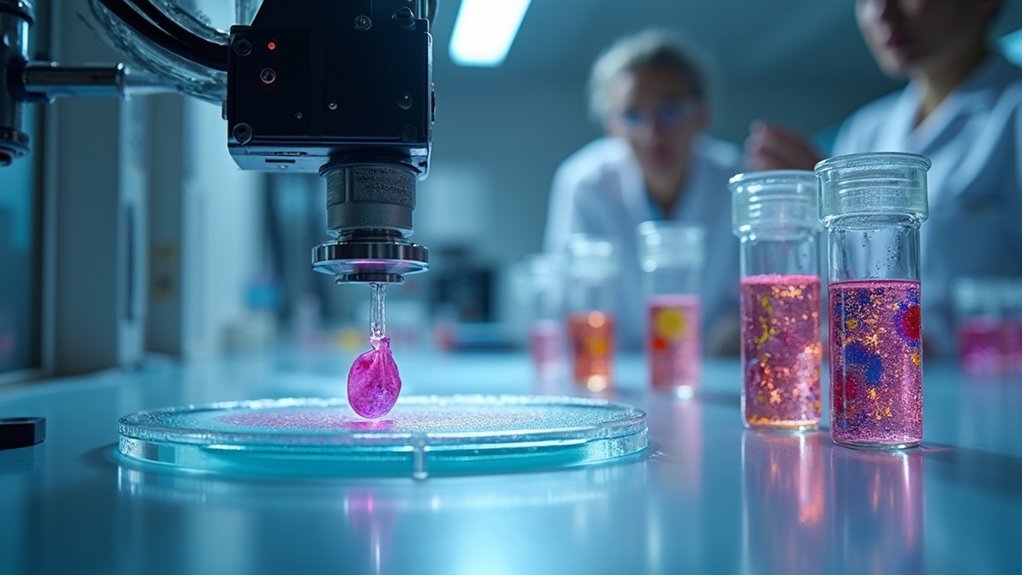
Advanced Direct Ink Writing (DIW) transforms your carefully selected bioink materials into precise, functional heart valve constructs through controlled layer-by-layer deposition.
This sophisticated bioprinting technology enables you to deposit PVA, Gelatin, and K-carrageenan bioinks with exceptional accuracy, creating complex geometries that replicate native heart valve anatomy. You’ll achieve high-resolution 3D constructs with mechanical properties matching natural tissue requirements.
DIW integrates seamlessly with computer-assisted design software, allowing you to customize valve geometries for individual patients.
Real-time parameter monitoring guarantees consistent results across different printing sessions. By eliminating support bath requirements, you’ll simplify your biofabrication workflow while enhancing mechanical stability.
This streamlined approach reduces variability and improves the overall quality of your tissue-engineered heart valves, making DIW essential for successful cardiac bioprinting applications.
Integrating Human Valve Interstitial Cells for Tissue Development
You’ll need to optimize your cell seeding methods to guarantee VICs distribute evenly throughout the bioprinted matrix and maintain viability during the printing process.
Understanding how these interstitial cells grow and proliferate within your 3D construct will determine whether your valve develops the proper mechanical strength and flexibility.
Success depends on creating the right conditions for tissue integration, where VICs can produce collagen and form the extracellular matrix that gives your bioprinted valve its structural integrity.
Cell Seeding Optimization Methods
When bioprinting heart valves, you’ll find that optimizing cell seeding techniques directly determines the success of your valve’s functionality and integration. Proper distribution of human valve interstitial cells guarantees effective tissue development throughout your bioprinted construct.
You should implement dynamic seeding by applying mechanical stimuli to enhance cell adhesion and proliferation. This approach markedly improves cell density and tissue quality.
Select scaffolds with tailored porosity and surface properties to facilitate better cell attachment and migration, promoting robust extracellular matrix formation.
Consider sequential seeding of different cell types within your hydrogel construct to create layered structures mimicking natural valve architecture.
Incorporate growth factors and biochemical cues during seeding to stimulate VICs, promoting collagen production and improving your bioprinted valve’s mechanical properties.
Interstitial Cell Growth Patterns
Since VICs constitute the primary cellular component of native heart valves, understanding their growth patterns becomes fundamental to successful bioprinting outcomes.
You’ll need to focus on spatial arrangement when integrating interstitial cells into your 3D bioprinted constructs, as heterogeneous distribution yields superior tissue development compared to uniform seeding.
Your VICs will enhance collagen type I production, directly improving mechanical strength of the engineered heart valves.
You’ll observe that proper spatial organization promotes natural tissue remodeling patterns that closely mimic native valve behavior.
Optimizing culture conditions becomes critical for maintaining VIC viability and functionality throughout the bioprinting process.
You should carefully control growth factors and environmental parameters to guarantee your interstitial cells contribute effectively to the overall tissue engineering success of your heart valve constructs.
Tissue Integration Success Factors
Although successful bioprinting depends on multiple variables, integrating human valve interstitial cells effectively requires you to master three critical success factors that determine whether your engineered tissue will achieve proper integration.
Your VICs must establish robust interactions with selected biomaterials to drive ideal tissue development. The bioink composition directly influences cellular behavior, affecting how well your bioprinted heart valves will function long-term. You’ll need to refine these material-cell interactions to guarantee successful tissue integration.
- Enhanced collagen production – VICs boost mechanical strength and durability through increased collagen synthesis within your engineered constructs.
- Improved physiological responses – Incorporating heterogeneous VIC populations enables better mimicry of native valve behavior and enhanced remodeling capabilities.
- Superior biocompatibility – VICs promote seamless integration between your bioprinted tissue and surrounding biological systems.
Creating Biomimetic Structures With Proper Hydrodynamic Properties
When you’re bioprinting heart valves, you’ll need to replicate the precise three-dimensional architecture of natural valves, including their leaflet curvature, commissure positioning, and annular dimensions.
You must design these structures to achieve laminar blood flow patterns that minimize turbulence and reduce hemolysis during the cardiac cycle.
Your success depends on balancing the geometric complexity with printability constraints while ensuring the valve opens and closes efficiently under physiological pressure gradients.
Mimicking Natural Valve Geometry
Three critical elements define successful heart valve bioprinting: precise geometric replication, optimal material selection, and hydrodynamic functionality.
When you mimic the structure of native valves, you’re creating anatomically accurate leaflets that guarantee optimal blood flow patterns. Advanced 3D bioprinting techniques like direct ink writing and stereolithography enable you to replicate complex valve architectures with remarkable precision.
Your bioprinted heart valves must incorporate biological hydrogels containing human valve interstitial cells to promote collagen production. This approach recreates the natural extracellular matrix essential for structural integrity.
You’ll need to carefully refine leaflet thickness and curvature to achieve proper hydrodynamic properties.
Key geometric considerations include:
- Leaflet curvature – Guarantees smooth opening and closing motions
- Commissural alignment – Maintains proper valve coaptation
- Annular dimensions – Matches patient-specific anatomical requirements
Optimizing Flow Dynamics
Computational fluid dynamics (CFD) simulations become your primary tool for predicting and enhancing blood flow patterns within bioprinted heart valves. You’ll use these simulations to guarantee your bioprinted valves replicate the hydrodynamics of healthy native valves, minimizing turbulence during cardiac cycles.
When designing your valve, incorporate anisotropic materials that improve mechanical properties, allowing leaflets to withstand dynamic pressures and shear forces. Focus on creating trileaflet valve structures that mirror natural aortic valves, as they demonstrate superior hemodynamic performance and reduced regurgitation.
You must maintain precise geometrical features and surface textures to achieve ideal blood flow dynamics. This attention to detail guarantees proper interaction with blood components, notably reducing thrombogenic risks and improving overall valve functionality.
Evaluating Mechanical Performance and Biological Functionality

After you’ve successfully bioprinted a heart valve, you must rigorously evaluate both its mechanical performance and biological functionality to confirm it can replace natural tissue effectively.
Your bioprinted heart valves need thorough testing to guarantee they’ll withstand physiological conditions. You’ll assess hydrodynamic behavior, durability under cyclic loading, and resistance to deformation. The Young’s modulus of your PVA-Gelatin-K-carrageenan bioink should closely match natural heart tissue for ideal functionality.
Rigorous mechanical testing ensures bioprinted heart valves match natural tissue properties and withstand the demanding cardiovascular environment.
- Test valve opening and closing using pulse duplicator simulations to verify natural heart valve mimicry
- Monitor biocompatibility through inflammatory response assessment and collagen development tracking over four weeks
- Evaluate calcification resistance compared to traditional mechanical valves for enhanced long-term viability
This rigorous evaluation confirms your tissue engineering efforts produce viable cardiac replacements.
Scaling Production for Clinical Viability and Long-term Durability
When shifting from laboratory prototypes to clinical implementation, you’ll need to establish standardized manufacturing protocols that maintain consistent biomaterial properties across large-scale production runs. Your bioprinted heart valves require precise control of viscosity and biocompatibility parameters to guarantee reliability across batches.
| Component | Requirement | Validation Method |
|---|---|---|
| Biomaterial Properties | Consistent viscosity control | Batch testing protocols |
| Printer Precision | High-resolution geometry | BIO X6 calibration |
| Quality Standards | Zero-defect production | Multi-point inspection |
You’ll achieve long-term durability by incorporating biocompatible materials like PVA/gelatin/carrageenan bioink, which resists calcification and thrombogenicity in preclinical studies. Implement robust quality assurance frameworks throughout production, and conduct continuous in vivo monitoring to validate performance for clinical applications.
Frequently Asked Questions
What Is the Life Expectancy of a Biological Heart Valve?
Your biological heart valve’ll typically last 10 to 20 years, though you might need replacement sooner if you’re younger. You’ll experience shorter lifespan than mechanical valves but won’t require lifelong blood thinners.
Can You Bioprint a Heart?
You can’t bioprint a complete functioning heart yet with current technology. However, you’re able to successfully bioprint individual heart components like valves and smaller tissue structures using specialized bioinks and advanced 3D printing techniques.
What Are the Disadvantages of Bioprosthetic Heart Valves?
You’ll face thrombogenicity risks, calcification buildup, limited durability requiring replacement every 10-15 years, lifelong anticoagulation needs, and multiple surgeries if you’re a pediatric patient since they don’t grow.
How to Decalcify Your Heart Valves?
You can’t directly decalcify your heart valves at home. Medical professionals use EDTA chelation therapy, specialized surgical procedures, or targeted pharmaceutical interventions. Consult your cardiologist for proper evaluation and treatment options.

Leave a Reply