You’ll start by selecting an appropriate collagen source like marine or recombinant options, then extract using acid or enzymatic methods. Purify the collagen through methacrylation processing while controlling viscosity between 100-1,000 Pa·s by adjusting concentration and adding alginate at a 3:1 ratio. Implement crosslinking strategies using photo or chemical methods for mechanical stability, then validate biocompatibility by integrating cells at 2×10⁶ cells/mL and monitoring viability. The complete methodology encompasses several critical optimization steps for successful bioink development.
Collagen Source Selection and Extraction Methods

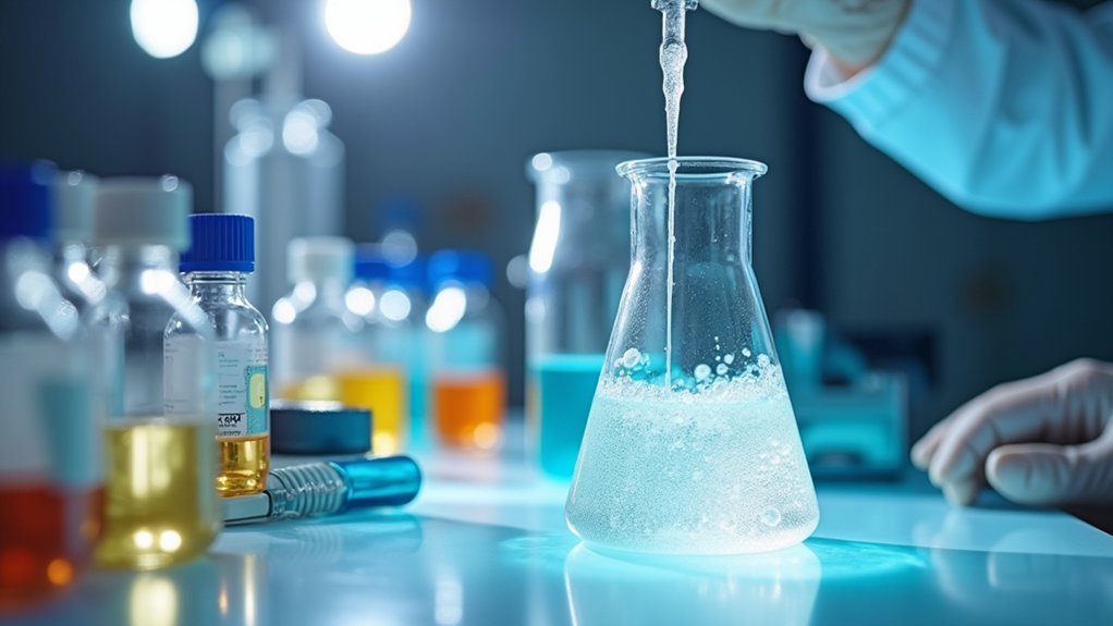
The foundation of successful collagen bioink creation begins with selecting the appropriate collagen source and extraction methods for your specific application.
You’ll find Type I collagen extracted from bovine, porcine, and marine species offers distinct advantages for biocompatibility in tissue engineering. Marine collagen presents lower immunogenicity and ethical sourcing benefits, making it ideal for patients with mammalian protein allergies.
Consider recombinant collagen production for engineered properties and consistent quality. Your extraction methods should utilize acid or enzyme-based processes targeting fibrous tissues like skin, tendons, and bones.
Implement rigorous quality control measures, as source tissue, extraction techniques, and processing conditions directly impact yield, structural integrity, and biological activity of your final bioink applications.
Purification and Processing Techniques for Bioprinting Applications
Once you’ve extracted your collagen, implementing effective purification techniques becomes critical for achieving bioink performance standards in 3D bioprinting applications.
You’ll need to remove non-collagenous proteins and fat through acid or enzymatic treatments, ensuring ideal biocompatibility for tissue engineering applications.
Processing techniques like methacrylation enhance your collagen bioinks’ printability and structural integrity.
Methacrylation processing significantly improves collagen bioink printability and maintains essential structural integrity for successful 3D bioprinting applications.
You can adjust viscosity by modifying collagen concentration and incorporating complementary biomaterials such as alginate to refine flow behavior during extrusion.
Crosslinking methods greatly impact your bioink’s performance.
You’ll choose between physical crosslinking (thermal or ionic) or chemical approaches using glutaraldehyde or genipin to improve mechanical properties and post-printing stability.
Establish rigorous quality control measures throughout purification and processing, as variations directly affect your bioink’s biocompatibility and overall performance in bioprinting applications.
Rheological Property Optimization and Viscosity Control

While successful collagen extraction and purification lay the foundation for quality bioinks, achieving ideal rheological properties determines whether your bioink will perform effectively during the printing process.
You’ll need to optimize viscosity between 100-1,000 Pa·s for successful extrusion and cell integration in tissue engineering applications.
Consider these key optimization strategies:
- Temperature control – Increase printing temperature to reduce viscosity while maintaining structural integrity
- Additive incorporation – Use sodium alginate at 3:1 ratio to enhance mechanical properties and printability
- Rheological assessment – Measure yield stress and flow index to optimize printing parameters
- Crosslinking methods – Implement ionic or photocrosslinking to control gelation time and structural stability
Fine-tuning these rheological properties guarantees your collagen bioinks achieve consistent strand diameters and successful tissue construct formation.
Crosslinking Strategies for Enhanced Mechanical Stability
Once you’ve optimized your collagen bioink’s rheological properties, you’ll need to enhance its mechanical stability through strategic crosslinking approaches.
You can choose between chemical crosslinking methods that create strong covalent bonds or physical crosslinking techniques that offer simpler, less toxic alternatives.
Each approach gives you different advantages in building structural integrity for your bioprinted constructs.
Chemical Crosslinking Methods
Although collagen bioinks offer excellent biocompatibility, their inherently weak mechanical properties often limit their effectiveness in tissue engineering applications.
You’ll need chemical crosslinking to enhance mechanical stability by forming covalent bonds between collagen molecules. These crosslinking strategies greatly improve your scaffold properties for demanding tissue engineering scenarios.
When implementing chemical crosslinking methods, consider these key approaches:
- Glutaraldehyde crosslinking – Creates strong covalent bonds but requires careful handling due to cytotoxicity concerns
- Genipin crosslinking – Offers biocompatible alternative with excellent mechanical enhancement
- Photo-crosslinking techniques – Provides spatial control using UV light with methacrylated collagen
- Multifunctional crosslinkers – Combines multiple strategies for superior performance
You can tailor degradation rates through controlled crosslinking, enabling precise release of bioactive factors while facilitating cell migration and tissue integration during healing processes.
Physical Crosslinking Techniques
Beyond chemical methods, physical crosslinking techniques offer you gentler alternatives that preserve collagen’s natural structure while strengthening your bioinks.
You can utilize ionic gelation with calcium ions to create stable hydrogel networks, greatly enhancing mechanical strength for tissue engineering applications.
Temperature-induced gelation allows your collagen solutions to shift into stable hydrogels at physiological temperatures, closely mimicking natural tissue conditions.
Freeze-thaw cycles provide another effective approach, forming porous structures that improve mechanical performance while facilitating nutrient transport.
You’ll find that incorporating natural polymers like alginate creates synergistic interactions that boost printability and crosslinking efficiency.
Additionally, mechanical forces during printing can reinforce your constructs’ structure, optimizing the overall mechanical properties of your collagen bioinks for demanding applications.
Cell Integration and Biocompatibility Testing Protocols

When developing collagen bioinks, you’ll need to establish rigorous protocols for evaluating how well cells integrate with your formulations and confirming they’re safe for biological applications.
Start by loading human pulmonary lung fibroblasts into your bioinks at 2 × 10⁶ cells/mL concentration for thorough cell integration assessment.
Load human pulmonary lung fibroblasts at 2 × 10⁶ cells/mL concentration to ensure comprehensive cell integration assessment in your bioink formulations.
Your biocompatibility testing protocol should include:
- Cell viability measurements using WST-1 assays to evaluate metabolic activity across different collagen ratios
- Mechanical properties evaluation including tensile strength and compressive modulus testing to guarantee structural integrity under physiological conditions
- Time-course studies monitoring proliferation and differentiation at Days 1, 5, and 10
- Statistical analysis using Kruskal-Wallis tests to compare cellular viability across varying formulations
These protocols guarantee your collagen bioinks support ideal cell growth without cytotoxic effects.
Quality Assessment and Characterization of Final Bioink Formulations
You’ll need to thoroughly evaluate your collagen bioink’s performance through extensive testing protocols that guarantee it meets the demanding requirements of bioprinting applications.
Start by conducting rheological property testing to measure viscosity and shear stress, which directly impact how smoothly your bioink will extrude through the printer nozzle.
Follow this with mechanical strength evaluation to assess compressive and tensile properties, then perform cellular viability assessment using relevant cell types to confirm your bioink supports proper cell function and survival.
Rheological Property Testing
Although creating your collagen bioink represents a significant milestone, you can’t determine its suitability for 3D bioprinting without thoroughly evaluating its rheological properties.
Rheological property testing evaluates your bioink’s flow behavior, directly impacting printability and scaffolds quality during the extrusion process.
You’ll need to assess these critical parameters:
- Viscosity measurements – Select appropriate needle sizes (200 µm for higher viscosity, 150 µm for lower viscosity formulations)
- Shear stress sweep tests – Determine mechanical properties and shear thinning behavior
- Temperature refinement – Maintain 25-45°C for ideal printing performance
- Herschel-Bulkley modeling – Calculate yield stress, consistency index, and flow index parameters
Remember that increased collagen concentration may decrease compressive modulus, affecting your bioink’s suitability for specific tissue engineering applications.
Mechanical Strength Evaluation
Mechanical strength evaluation represents the final critical step in validating your collagen bioink’s performance for tissue engineering applications. You’ll conduct compression testing to assess your bioink’s ability to withstand axial loads, measuring compressive modulus values. Note that higher collagen concentrations may paradoxically decrease mechanical strength. Tensile testing determines bond strength between printed layers, calculating Young’s modulus from stress-strain curves.
| Test Type | Property Measured | Key Insight |
|---|---|---|
| Compression | Compressive Modulus | Higher collagen may reduce stiffness |
| Tensile | Young’s Modulus/UTS | Indicates structural integrity |
| Rheological | Viscosity | Affects printability and scaffold shape |
You’ll perform statistical analysis using ANOVA or Kruskal-Wallis tests to compare mechanical properties across formulations, ensuring your bioink meets specific tissue engineering requirements while maintaining ideal printability characteristics.
Cellular Viability Assessment
Following mechanical characterization, cellular viability assessment validates your bioink’s biocompatibility through systematic evaluation of cell survival and proliferation within printed constructs.
You’ll culture human pulmonary lung fibroblasts within your collagen bioinks to determine ideal alginate-collagen ratios. Expect initial cell population decreases during the first three days, followed by recovery phases where specific formulations support enhanced proliferation.
Key assessment protocols include:
- Testing various bioink formulations using 3:1 and 2:1 alginate-collagen ratios for consistent viability increases after Day 5
- Conducting nonparametric Kruskal-Wallis statistical analysis to identify significant differences in cell proliferation
- Monitoring extended culture periods to detect decreased viability in higher collagen concentrations like 1:1 ratios
- Maintaining refined balance between components to guarantee successful tissue scaffolds for tissue engineering applications
This systematic approach guarantees your final formulations demonstrate proven biocompatibility for effective tissue engineering.
Frequently Asked Questions
How to Make Collagen Bioink?
You’ll dissolve type I collagen in acetic acid at 4% w/v concentration. Mix with sodium alginate in a 3:1 ratio, then methacrylate for crosslinking enhancement and ideal printability.
How to Create Bioink?
You’ll start by selecting your base biomaterial, then dissolve it in appropriate buffer solutions. Add crosslinking agents and stabilizers, adjust viscosity for printability, then characterize rheological properties before testing compatibility.
What Is the Formulation of Bioink?
You’ll typically combine natural polymers like collagen with synthetic materials such as alginate in specific ratios, often 3:1 sodium alginate to collagen, dissolved in PBS buffer for ideal printability.
What Is the Application of Collagen in Tissue Engineering?
You’ll find collagen’s essential for tissue engineering because it provides biocompatible scaffolds that support cell adhesion and growth. You can use it for heart, skin, cartilage, and vascular tissues, enhancing mechanical properties.


Leave a Reply