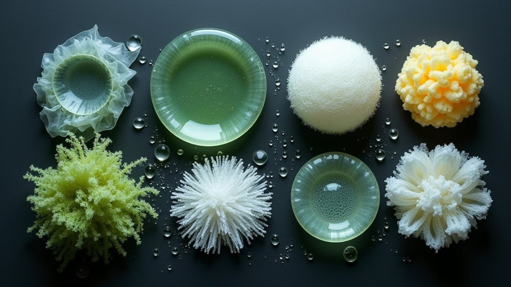
You’ll find seven exceptional biocompatible materials for tissue scaffold printing: Polycaprolactone (PCL) offers flexible structures with 60°C melting points, while Polylactic Acid (PLA) provides robust mechanical performance from sustainable sources. Hydroxyapatite mimics natural bone composition, and collagen-based constructs deliver mechanical strength through triple helix structures. PLGA composites feature customizable degradation timelines, alginate hydrogels support soft tissue applications with high water retention, and chitosan networks enhance cell adhesion. These materials collectively reveal advanced regenerative medicine possibilities that await your exploration.
Polycaprolactone (PCL) for Flexible Tissue Engineering Applications
Flexibility defines the essence of successful tissue engineering, and polycaprolactone (PCL) emerges as a standout biodegradable polymer that delivers exactly this characteristic.
Polycaprolactone stands as tissue engineering’s flexible foundation, delivering the adaptability essential for successful regenerative medicine applications.
You’ll find PCL’s low melting point of 60°C makes it perfect for flexible applications while maintaining impressive mechanical properties with 40 MPa tensile strength and 300-600 MPa Young’s modulus that mimics soft tissues.
You can control PCL’s degradation rate through molecular weight adjustments, ensuring scaffolds break down over 2-3 years to match tissue regeneration timelines.
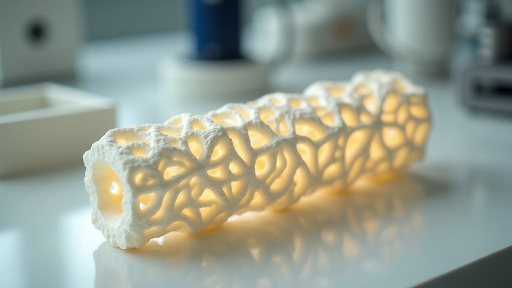
When you use electrospinning techniques, you’ll create nanofibrous structures with high surface area-to-volume ratios that enhance cell attachment and proliferation.
The biocompatible nature of this biocompatible material shows minimal inflammatory response, making it ideal for tissue engineering applications.
Polylactic Acid (PLA) Biodegradable Polymer Scaffolds
When you’re seeking a biodegradable polymer that combines exceptional printability with robust mechanical performance, polylactic acid (PLA) stands as your go-to solution for tissue scaffold applications. This biocompatible material excels in 3D printing techniques, creating complex structures that mimic the natural extracellular matrix while supporting ideal cell attachment and growth.
| Property | Specification | Benefit |
|---|---|---|
| Degradation Rate | Months to years | Matches tissue regeneration timelines |
| Source Material | Corn starch, sugarcane | Sustainable renewable resources |
| Cellular Response | Enhanced osteoblast function | Promotes bone regeneration |
| Processing Method | 3D printable | Complex scaffold geometries |
PLA’s tunable degradation rate, determined by molecular weight and composition, guarantees your scaffolds dissolve as new tissue forms. When combined with bioactive substances or stem cells, these biodegradable polymers greatly enhance bone regeneration outcomes in tissue scaffold printing applications.
Hydroxyapatite Ceramic Materials for Bone Regeneration
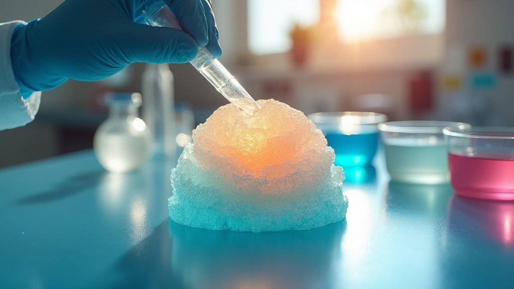
Although synthetic materials often fall short of replicating bone’s natural properties, hydroxyapatite (HA) ceramic materials bridge this gap by mimicking the primary inorganic component found in your body’s bone tissue.
These synthetic biomaterials offer exceptional biocompatibility and promote osteoblast proliferation, making them ideal for bone regeneration applications.
When you incorporate HA into composite scaffolds, you’ll greatly enhance both mechanical properties and bioactivity compared to pure polymers.
3D printing technology allows precise control over scaffold architecture, optimizing pore distribution for cellular infiltration and nutrient flow.
You can produce synthetic hydroxyapatite through sol-gel and precipitation methods, achieving crystalline structures that closely match natural bone minerals.
These scaffolds chemically bond with surrounding tissue, accelerating new bone formation and providing superior outcomes in tissue engineering applications.
Collagen-Based Natural Polymer Constructs
When you’re working with collagen-based constructs for tissue scaffolds, you’ll need to understand how collagen’s triple helix structure and natural cross-linking sites directly impact scaffold performance and cellular integration.
You can process collagen through various bioprinting methods including extrusion-based printing, inkjet deposition, and laser-assisted techniques, each offering different advantages for controlling fiber alignment and porosity.
You’ll often need to enhance collagen’s inherently weak mechanical properties through cross-linking agents, polymer blending, or composite reinforcement to meet the structural demands of load-bearing tissue applications.
Collagen Structure Properties
As you explore biocompatible materials for tissue scaffold printing, collagen emerges as the most promising natural polymer due to its unique structural properties and biological compatibility.
You’ll find that collagen’s triple helix structure provides exceptional mechanical strength while maintaining flexibility essential for tissue applications. Its remarkable biocompatibility stems from its similarity to the natural extracellular matrix (ECM), promoting optimal cell adhesion and proliferation.
When you design collagen scaffolds, you can create porous scaffolds that mimic native tissue architecture.
You’ll appreciate how collagen’s fibril-forming ability enables controlled porosity for nutrient transport. Additionally, you can incorporate bioactive molecules to enhance tissue regeneration responses.
The controllable biodegradation rate through cross-linking methods allows you to match scaffold breakdown with natural healing processes, making collagen ideal for regenerative applications.
Bioprinting Processing Methods
Since collagen’s structural properties make it ideal for tissue engineering, you’ll need to understand the specific bioprinting processing methods that maximize its potential in scaffold construction.
These biocompatible scaffolds require precise layer-by-layer deposition techniques that control architecture while maintaining collagen’s natural extracellular matrix characteristics.
Key processing enhancement strategies include:
- Concentration adjustment – Varying collagen concentrations to achieve target mechanical properties
- Cross-linking control – Implementing specific cross-linking methods for desired biodegradable rates
- Growth factor integration – Incorporating bioactive molecules directly into the matrix during printing
- Cell incorporation – Adding live cells during deposition to enhance cell adhesion and proliferation
Advanced bioprinting methods now enable vascular network formation within collagen constructs, ensuring proper nutrient transport.
You’ll achieve superior results by matching processing parameters to your specific tissue engineering application requirements.
Mechanical Strength Enhancement
While collagen’s inherent biocompatibility makes it an excellent foundation for tissue scaffolds, you’ll often encounter limitations in mechanical strength that require strategic enhancement approaches.
You can considerably improve tensile strength through cross-linking agents like glutaraldehyde or genipin, which stabilize the structure while preserving cellular compatibility.
Electrospun collagen creates nanofibrous scaffolds with enhanced surface area and porosity, dramatically improving mechanical properties and facilitating nutrient transport.
You’ll achieve even better results by developing composite scaffolds that combine collagen with materials like hydroxyapatite or chitosan. These combinations boost osteoconductivity and mechanical performance, making them ideal for bone tissue regeneration.
Research shows you can achieve compressive strengths exceeding 10 MPa, matching native cartilage properties for effective tissue repair applications.
Poly(Lactic-co-Glycolic Acid) (PLGA) Composite Scaffolds
When you’re selecting PLGA for tissue scaffold printing, you’ll find its degradation timeline can be precisely controlled by adjusting the lactic acid to glycolic acid ratio, allowing customization from weeks to years based on your specific tissue application.
You can greatly enhance PLGA’s mechanical properties and bioactivity by creating composites with natural polymers like collagen or chitosan, maintaining biodegradability while boosting scaffold performance.
This composite approach lets you combine PLGA’s tunable degradation with the superior biological properties of natural materials, creating scaffolds that better support cell growth and tissue integration.
PLGA Degradation Properties
The degradation timeline of PLGA scaffolds serves as a critical engineering parameter that you can precisely control to match your specific tissue regeneration requirements.
This biodegradable polymer breaks down through hydrolysis, producing lactic and glycolic acids that your body naturally metabolizes without toxic accumulation.
You can engineer the degradation rate by adjusting:
- Lactic to glycolic acid ratios in the polymer composition
- Molecular weight of the PLGA material
- Scaffold design porosity and surface area
- Addition of specific polymer blends or additives
The mechanical properties gradually decrease as degradation progresses, allowing natural tissue to replace the scaffold structure.
This synchronized process guarantees ideal cell adhesion and biocompatibility throughout tissue engineering applications, making PLGA ideal for bone, cartilage, and skin regeneration where controlled support timing is essential.
Composite Material Integration
Although pure PLGA scaffolds offer excellent biocompatibility and controllable degradation, you’ll achieve superior tissue engineering outcomes by integrating composite materials that address PLGA’s inherent limitations.
By incorporating bioactive glass into your PLGA scaffolds, you’ll enhance osteoconductivity and mechanical properties while maintaining ideal degradation rates. These composite scaffolds provide controlled release of bioactive compounds that accelerate tissue regeneration.
You can fabricate these biocompatible composite structures using electrospinning or 3D printing techniques, creating versatile architectures that mimic natural extracellular matrix patterns.
When you optimize porosity in your PLGA composite scaffolds, you’ll markedly improve cell attachment, proliferation, and differentiation. This integration approach makes composite PLGA scaffolds particularly effective for bone and cartilage regeneration applications in modern tissue engineering.
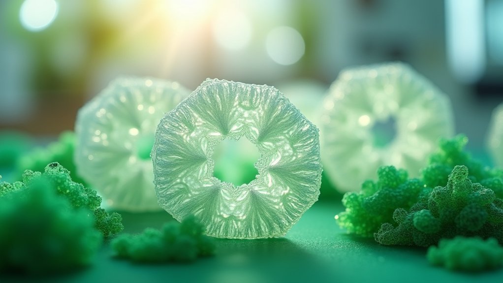
Alginate Hydrogel Matrices for Soft Tissue Applications
Among nature’s most versatile biomaterials, alginate hydrogels have emerged as leading candidates for soft tissue scaffold applications. You’ll find these seaweed-derived materials exceptionally biocompatible, supporting enhanced cell viability through their hydrophilic networks.
When you introduce calcium ions, alginate undergoes ionic crosslinking, creating tunable porous structures that facilitate nutrient diffusion and cell migration.
Key advantages you’ll experience with alginate scaffolds include:
- Easy modification with bioactive molecules like growth factors
- High water retention mimicking physiological tissue environments
- Seamless integration with advanced 3D printing techniques
- Customizable architectures for specific tissue applications
You can enhance tissue regeneration by incorporating peptides or growth factors directly into the matrix.
The material’s versatility allows you to create complex geometries through bioprinting, making alginate hydrogels ideal for personalized soft tissue engineering solutions.
Chitosan Biopolymer Networks for Enhanced Cell Adhesion
When you’re seeking superior cell adhesion properties in tissue scaffolds, chitosan biopolymer networks deliver exceptional performance through their unique cationic structure. This biocompatible biopolymer’s positive charge creates strong interactions with negatively charged cell membranes, promoting enhanced integration and proliferation.
| Property | Chitosan Benefit | Application Impact |
|---|---|---|
| Cell Adhesion | Cationic surface charge | Enhanced tissue integration |
| Mechanical Properties | Composite networks formation | Improved scaffold durability |
| Nutrient Diffusion | Porous architecture | Sustained cell viability |
| Bioactive Factors | Natural healing stimulants | Accelerated regeneration |
| Biodegradability | Complete tissue absorption | Safe regenerative medicine |
You can combine chitosan with materials like gelatin to create composite networks that enhance mechanical properties while maintaining excellent nutrient diffusion. These bioactive factors naturally stimulate cell migration, making chitosan ideal for regenerative medicine applications.
Frequently Asked Questions
What Biomaterials Are Used in Tissue Engineering Scaffolds?
You’ll find biodegradable polymers like PCL, PLA, and PGA combined with natural materials including collagen, chitosan, and alginate. You can also use bioactive ceramics such as hydroxyapatite and composite blends for enhanced performance.
What Materials Are Used in Tissue Scaffolds?
You’ll find biodegradable polymers like PCL, PLA, and PLGA commonly used in tissue scaffolds. Natural materials including collagen, chitosan, and alginate provide excellent biocompatibility, while ceramics like hydroxyapatite enhance bone formation.
What Synthetic Biodegradable Product Is Used as a Tissue Engineered Scaffold?
You’ll find polycaprolactone, polylactic acid, and poly(lactic-co-glycolic acid) are commonly used synthetic biodegradable products for tissue-engineered scaffolds. They’re chosen because they provide excellent biocompatibility, mechanical strength, and controllable degradation rates.
What Are Biocompatible Scaffolds?
You’ll find biocompatible scaffolds are specially designed materials that support your body’s tissue regeneration without causing harmful reactions. They provide structural frameworks where your cells can attach, grow, and form new healthy tissues.


Leave a Reply