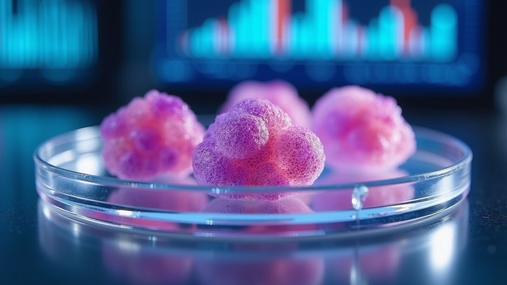
You’re seeing cancer research transform as 3D bioprinted tumor models replace traditional 2D cultures with sophisticated microenvironments that mirror real cancer conditions. These models incorporate patient-derived cells within complex structures, enabling cancer-immune cell interactions that dramatically improve drug response predictions. You’ll benefit from personalized medicine approaches where your unique tumor characteristics guide tailored therapies through high-throughput screening. This technology accelerates drug discovery while reducing animal testing, though current limitations in replicating temporal dynamics present ongoing challenges that emerging innovations continue to address.
How 3D Bioprinting Transforms Cancer Research Models

While traditional 2D cancer models have dominated research for decades, 3D bioprinting now transforms how scientists study tumor behavior by creating sophisticated microenvironments that mirror real cancer conditions.
You’ll find these bioprinted cancer research models incorporate patient-derived cells within complex extracellular matrix structures, enabling interactions between cancer cells and immune cells that 2D cultures can’t replicate.
This advancement dramatically improves predictive accuracy for drug responses compared to conventional methods.
You can now conduct high-throughput drug screening with greater confidence, as these models better predict how treatments will perform in actual patients.
The technology accelerates personalized medicine by allowing you to test specific drug responses using individual patient cells, streamlining the shift from in vitro testing to clinical applications while reducing development costs.
Advanced Hydrogels and Bioinks for Tumor Recreation
Since tumor microenvironments possess unique mechanical and biochemical properties, advanced hydrogels like GelMA and alginate have become essential bioinks that you can fine-tune to recreate these complex conditions.
These bioprinting materials offer excellent biocompatibility while maintaining cell viability and mimicking the extracellular matrix found in tumors.
You can adjust mechanical properties and degradation rates through careful polymer selection and crosslinking methods, enabling precise tumor microenvironment recreation.
Hydrogels accommodate multiple cell types and growth factors, allowing you to study complex tumor-stromal interactions effectively.
Recent hybrid bioinks combine natural and synthetic polymers, enhancing mechanical strength and printability.
Additionally, gradient hydrogels provide spatial control over biochemical factor distribution, giving you realistic conditions to study cell migration and tumor behavior patterns.
Personalized Medicine Through Patient-Specific Tumor Models

As precision medicine continues transforming cancer treatment, you can now harness patient-derived cells to create personalized 3D bioprinted tumor models that predict individual therapeutic responses with unprecedented accuracy.
These models replicate each patient’s unique tumor microenvironment, enabling precise drug dosage determination and enhanced treatment efficacy. You’ll discover cancer susceptibility patterns and therapy resistance mechanisms specific to individual patients, revolutionizing treatment strategies.
Through high-throughput drug screening on these personalized platforms, you can identify ideal tailored therapies faster than ever before.
The models effectively simulate tumor heterogeneity, providing superior predictive power compared to traditional 2D cultures. By integrating patient-specific characteristics into bioprinted tumor models, you’re advancing toward truly individualized cancer care that maximizes therapeutic outcomes while minimizing adverse effects.
Drug Discovery Acceleration With Bioprinted Cancer Tissues
Though traditional drug discovery relies heavily on oversimplified 2D cell cultures that fail to capture the complexity of human tumors, you can now accelerate pharmaceutical development using bioprinted cancer tissues that faithfully recreate the three-dimensional tumor microenvironment.
These bioprinted tumor models enable high-throughput drug screening, facilitating faster shifts to clinical applications. You’ll find that 3D bioprinted cancer tissues replicate essential cell-matrix interactions that greatly influence drug diffusion and treatment sensitivity.
When you use patient-derived cells to create personalized medicine platforms, you can predict individual responses more accurately than conventional methods. This approach reveals important differences—drugs like cisplatin show reduced efficacy in 3D models compared to 2D cultures, providing more reliable predictions for therapeutic outcomes.
Reducing Animal Testing While Improving Research Accuracy

While conventional animal models have long served as the bridge between laboratory research and human clinical trials, bioprinted tumor models now offer you a revolutionary alternative that simultaneously reduces ethical concerns and improves research accuracy.
These 3D bioprinting technologies create complex tissue structures that better replicate the tumor microenvironment compared to traditional 2D cultures. You’ll find that bioprinted models using patient-derived cells provide more relevant research findings for personalized cancer therapies.
This enhanced accuracy addresses the critical issue where 90% of drugs fail after preclinical studies due to inadequate testing models. By enabling high-throughput screening and streamlining drug development timelines, you can greatly reduce animal testing while achieving more predictive outcomes that better reflect human responses.
Current Limitations and Emerging Technologies in Bioprinted Oncology
Although bioprinted tumor models represent a significant advancement in cancer research, you’ll encounter several critical limitations that currently restrict their full potential.
Despite revolutionary progress in bioprinted cancer models, researchers must navigate significant technical barriers that currently limit their clinical translation potential.
These bioprinted models struggle to replicate the tumor microenvironment’s temporal dynamics and complex interactions essential for accurate drug development. You’ll also face scaling challenges when attempting industrial applications and maintaining long-term construct stability.
However, emerging technologies offer promising solutions. Advanced 3D bioprinting techniques, including extrusion-based methods and droplet-based approaches, enhance model accuracy and reproducibility for studying drug responses.
You’ll benefit from ongoing research into hybrid bioinks and multimaterial printing, which improve construct complexity and functionality. These innovations facilitate personalized therapy development and more effective drug screening, ultimately addressing interspecies differences that limit traditional animal models in human physiology representation.
Frequently Asked Questions
What Is the New Cancer Treatment That Melts Tumors?
You’ll receive tumor melting treatment that combines drugs inducing cancer cell death while boosting your immune system’s ability to destroy tumors. It’s delivered directly into tumors, minimizing side effects.
What Has Been Successfully Bioprinted?
You’ve seen researchers successfully bioprint 3D tumor models for glioblastoma, breast, lung, and colorectal cancers using patient-derived cells and hydrogels, plus advanced glioblastoma-on-a-chip systems with hypoxic cores.
What Is 3D Bioprinting of Tumor Models for Cancer Research?
You’re using layer-by-layer cell deposition to create realistic tumor environments that mimic cancer’s complexity, enabling better drug testing and personalized treatment development compared to traditional flat cell cultures.
What Are the Recent Advances in 3D Printing for in Vitro Cancer Models?
You’ll find exciting advances including improved extrusion bioprinting techniques, enhanced hydrogel properties for better biocompatibility, and patient-specific tumor constructs that enable personalized drug screening and high-throughput therapeutic evaluations.


Leave a Reply