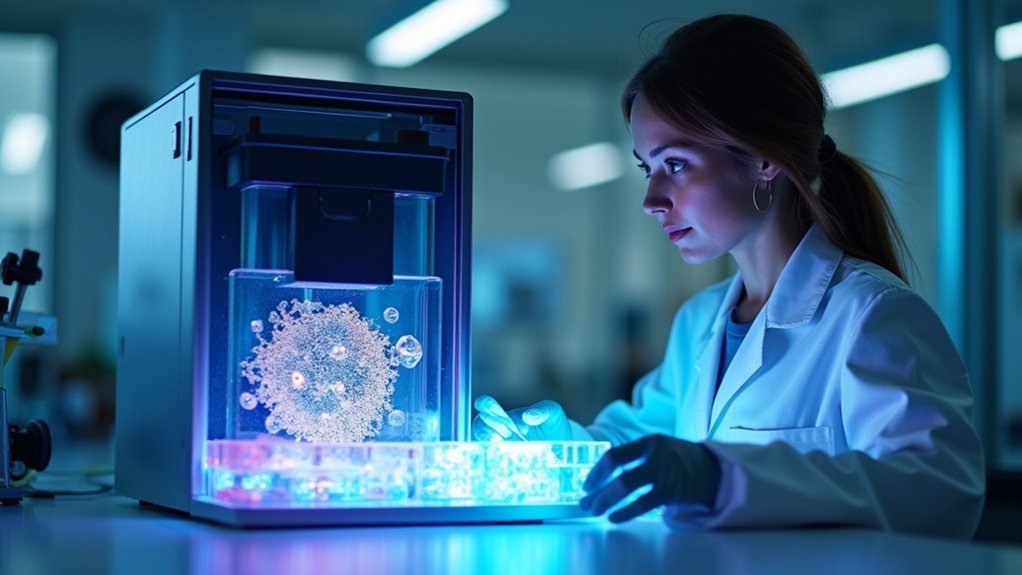
You’ll master pancreatic islet bioprinting by optimizing PINE bioink with 74.8% matrisome proteins and 0.2 mg/mL basement membrane concentrations. Keep printing pressures under 30 kPa while using low-viscosity hydrogels to preserve cell viability. Design microvascular channels that mimic native pancreatic architecture for proper nutrient flow. Implement staged differentiation protocols monitoring PDX1 and NKX6.1 markers throughout maturation. Validate functionality through glucose-stimulated insulin secretion testing at 16.7 mM concentrations. These foundational techniques reveal advanced bioprinting strategies for therapeutic applications.
Optimize PINE Bioink Composition With Pancreas-Derived ECM and Basement Membrane Proteins
When bioprinting pancreatic islet cells, you’ll achieve superior results by optimizing your PINE bioink composition with pancreas-derived extracellular matrix (pdECM) and basement membrane proteins.
The pdECM contains 74.8% matrisome proteins, providing pancreatic-specific components essential for stem cell differentiation. You’ll want to maintain basement membrane protein concentrations around 0.2 mg/mL to maximize PDX1 and CHGA expression, promoting functional β cells development.
The critical glycoproteins and proteoglycans in your bioink composition modulate signaling pathways that enhance glucoregulatory functions. By fine-tuning these components, you’ll create specialized niches that maintain structural integrity of islet aggregates while improving their physiological performance, ultimately leading to enhanced insulin secretion responses to glucose stimuli.
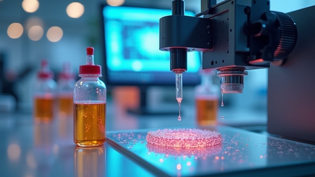
Control Printing Parameters to Minimize Mechanical Stress on Delicate Islet Cells
Your carefully optimized bioink composition sets the foundation, but controlling printing parameters becomes equally important to protect the fragile architecture of pancreatic islet cells during extrusion.
Keep bioprinting pressures under 30 kPa to maintain cell viability, as excessive pressure dramatically reduces cell survival rates.
Maintain bioprinting pressures below 30 kPa to preserve pancreatic islet cell viability and prevent catastrophic drops in survival rates.
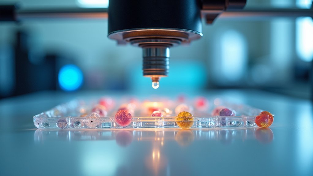
Select nozzle sizes carefully—larger ones create harmful shear stress, while smaller sizes improve precision but risk blockages.
Reduce printing speed to give islet cells time to adapt during the extrusion process.
Choose low-viscosity hydrogels that minimize mechanical stress on delicate cells.
Regular bioprinter calibration and monitoring of your bioink’s rheological properties guarantee consistent conditions that protect islet cell integrity throughout the printing procedure.
Design Vascularized Architecture for Enhanced Nutrient Flow and Glucose Sensing

Although protecting cells during printing is essential, creating vascularized networks within your bioprinted constructs determines whether islet cells will thrive long-term or fail due to inadequate oxygen and nutrient supply.
You’ll need sophisticated vascularization strategies that incorporate microvascular channels throughout your pancreatic islets to guarantee proper nutrient flow and glucose sensing capabilities.
Modern bioprinting technology allows you to design geometric patterns that create ideal niches for enhanced cell survival and metabolic activity.
Unlike traditional hydrogel-based encapsulation methods that often cause hypoxia, vascularized islet constructs demonstrate considerably improved glucose responsiveness.
You should focus on mimicking native pancreatic architecture by positioning vascular networks strategically to facilitate synchronized interactions between islet cells and blood vessels, assuring your bioprinted tissues maintain functionality.
Implement Staged Differentiation Protocols for Stem Cell-Derived Beta Cell Maturation
Since successful bioprinting relies on functional cells rather than just structural integrity, you’ll need to master staged differentiation protocols that transform pluripotent stem cells into mature, insulin-producing cells.
Monitor key markers like PDX1 and NKX6.1 throughout your stem cell-derived beta cell maturation process. You’ll want to optimize basement membrane protein concentrations to enhance these markers’ expression.
Monitor PDX1 and NKX6.1 markers continuously during beta cell maturation while optimizing basement membrane proteins to enhance expression levels.
Control your extracellular matrix components and growth factors carefully during each stage. This approach considerably boosts hormone production, with insulin, glucagon, and somatostatin levels rising dramatically compared to undifferentiated cells.
Properly matured islets respond to elevated glucose levels with increased insulin secretion, proving their viability for therapeutic applications. Use immunostaining techniques to confirm successful conversion and validate your differentiation protocol’s effectiveness.
Validate Functional Performance Through Glucose-Stimulated Insulin Secretion Testing
After you’ve successfully differentiated your stem cells into mature beta cells, glucose-stimulated insulin secretion (GSIS) testing becomes your primary validation method for confirming functional performance.
You’ll need to expose your bioprinted pancreatic islet cells to 16.7 mM glucose concentration to trigger robust insulin secretion responses. Monitor calcium intensity profiles during testing, as pronounced Ca²⁺ responses indicate effective insulin release mechanisms and enhanced metabolic activity.
Don’t overlook the importance of extracellular matrix components in your bioink formulation—specialized matrices like PINE markedly improve GSIS testing outcomes by enhancing glucoregulatory functions.
Your islet-like cellular aggregates should demonstrate markedly increased insulin secretion levels under high glucose stimuli, confirming functional maturation. Consistent GSIS testing validates that your bioprinted islets respond similarly to native pancreatic tissue.
Frequently Asked Questions
Can You 3D Print a Pancreas?
You can’t 3D print a complete functional pancreas yet. However, you can bioprint pancreatic islet structures that maintain insulin secretion with up to 90% cell viability, offering promising diabetes treatment possibilities.
How to Regenerate Islet Cells?
You’ll regenerate islet cells by creating supportive ECM environments using specialized bioinks, incorporating growth factors for stem cell differentiation, ensuring proper vascularization for nutrient supply, and continuously monitoring insulin secretion and metabolic function.
What Is the Success Rate of Pancreatic Islet Cell Transplant?
You’ll find pancreatic islet transplant success rates reach 40-50% for insulin independence after one year, but they drop dramatically to only 10-20% after five years due to rejection and inadequate vascularization.
How Can I Regenerate My Pancreas Beta Cells Naturally?
You can naturally support beta cell regeneration by eating antioxidant-rich foods, exercising regularly, maintaining healthy weight, managing blood sugar levels, and considering supplements like omega-3s, vitamin D, berberine, and curcumin.


Leave a Reply