You’ll succeed in bioprinted cartilage repair by optimizing bioink formulations with alginate and collagen, incorporating growth factors like TGF-β1 and BMP-2 for chondrogenic differentiation, and selecting appropriate cell types such as iPSCs. Design multi-material scaffolds with varying stiffness zones, utilize advanced CT and MRI imaging for precise printing guidance, and monitor cellular health throughout the process. Target specific mechanical properties like 1.8 MPa aggregate modulus for articular cartilage while ensuring proper matrix production and scaffold integration for ideal regenerative outcomes ahead.
Optimize Bioink Formulation for Enhanced Biocompatibility
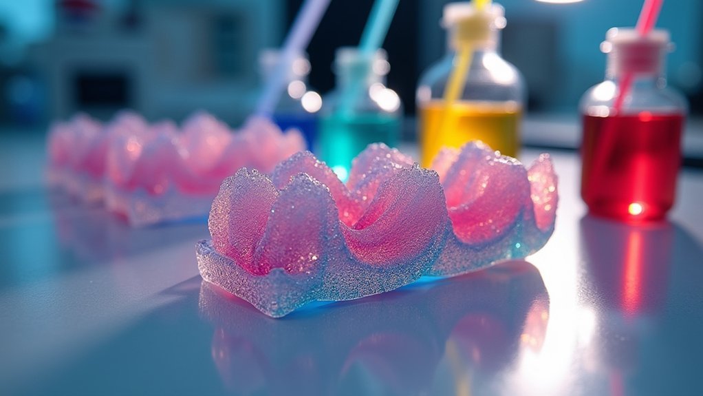
When developing bioinks for cartilage repair, you’ll want to prioritize natural biomaterials like alginate and collagen as your foundational components.
These biocompatible materials effectively mimic the extracellular matrix, providing essential structural support that enhances cell adhesion and proliferation. You should incorporate growth factors like TGF-β1 and BMP-2 directly into your bioink formulations to stimulate chondrogenic differentiation and matrix secretion.
Focus on refining your bioinks’ rheological properties to guarantee smooth dispensing through printing nozzles while maintaining cell viability.
Consider developing composite bioinks that blend natural and synthetic materials, achieving ideal mechanical properties for cartilage regeneration.
Always conduct thorough in vitro testing to assess cell viability, proliferation rates, and matrix production before proceeding to clinical applications.
Select Appropriate Cell Types for Chondrogenic Differentiation
Your bioink formulation sets the foundation, but selecting the right cell types determines whether your printed construct will successfully differentiate into functional cartilage tissue.
Choose induced pluripotent stem cells (iPSCs) for their exceptional ability to generate abundant chondrocytes while maintaining pluripotency within bioinks. You’ll enhance chondrogenic differentiation by co-culturing iPSCs with irradiated mature chondrocytes, creating an ideal cellular environment.
iPSCs offer superior chondrocyte generation while preserving pluripotency, with co-culturing alongside irradiated mature chondrocytes optimizing the differentiation environment.
Incorporate essential growth factors like TGFβ1, TGFβ3, GDF5, and BMP2 to promote cartilage matrix formation, specifically collagen types II, IX, and XI.
Monitor pluripotency markers such as OCT4 before and after bioprinting to guarantee your cells retain differentiation potential.
Remember that your mechanical environment greatly influences cell behavior, so consider how your selected cell types interact with bioink components to achieve successful cartilage repair outcomes.
Incorporate Growth Factors to Accelerate Tissue Regeneration
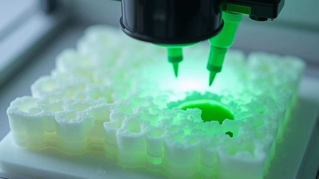
Although cell selection provides the cellular foundation, strategically incorporating growth factors into your bioink formulation will greatly accelerate chondrogenic differentiation and matrix production.
You’ll want to include TGFβ1, TGFβ3, GDF5, and BMP2 in your bioprinting protocols to enhance cartilage regeneration effectively. These growth factors promote expression of essential collagen types II, IX, and XI, plus aggrecan—critical components for functional cartilage formation.
Focus on controlled release mechanisms within your bioinks to maintain chondrocyte viability and stimulate proliferation.
Combining multiple bioactive factors greatly increases glycosaminoglycan production, which is fundamental for cartilage function. This approach mimics the natural healing environment, ensuring your tissue repair outcomes are optimized.
Proper growth factor integration transforms basic cellular scaffolds into powerful regenerative constructs.
Design Multi-Material Scaffolds for Complex Cartilage Structures
When designing multi-material scaffolds, you’ll need to strategically combine natural polymers like alginate and collagen with synthetic materials to achieve ideal performance for complex cartilage structures.
You must focus on creating seamless interfaces between different materials that maintain structural integrity while supporting distinct cellular environments.
Your scaffold design should match the mechanical properties of native cartilage by carefully balancing stiffness and flexibility across different zones to replicate the heterogeneous nature of cartilage tissue.
Material Selection Strategies
The foundation of successful cartilage bioprinting lies in selecting biomaterials that replicate the intricate mechanical and biological properties of native tissue. Your material selection strategy should combine type II collagen for load-bearing capabilities with hyaluronic acid to enhance hydrophilicity and cell adhesion.
You’ll want to incorporate nanocomposite materials into your scaffold design to boost mechanical strength and durability, which studies show increases type II collagen expression in cultured chondrocytes.
Consider using bioinks that blend natural biomaterials like alginate and silk fibroin for ideal degradation rates and tissue integration.
Advanced bioprinting techniques, including gradient printing, enable spatially controlled material deposition. Strategic layering creates distinct microenvironments that promote varying cellular behaviors, ultimately enhancing your scaffold’s regenerative potential while simulating native cartilage’s heterogeneous composition.
Interface Integration Techniques
Since cartilage repair demands scaffolds that can seamlessly integrate multiple tissue zones, you’ll need to master interface integration techniques that create stable connections between different biomaterial regions.
Focus on coaxial bioprinting methods that create core-shell structures, enabling you to combine materials like GelMA and PEGDA for enhanced mesenchymal stem cells differentiation into cartilage-like tissues.
You must carefully coordinate degradation rates between biomaterials to prevent premature scaffold failure.
Design your interface connections to match cartilage’s varying stiffness requirements—this directly influences chondrocyte behavior and matrix production.
Implement controlled growth factor release at interfaces to promote cell interactions essential for tissue integration.
Your bioprinting parameters should guarantee mechanical stability while maintaining ideal microenvironments for regeneration across different scaffold zones.
Mechanical Property Matching
Although cartilage exhibits complex mechanical properties with an aggregate modulus of 1.8 MPa and directional tensile strength reaching dozens of MPa in meniscal tissue, you can engineer multi-material scaffolds that replicate these demanding requirements through strategic biomaterial combinations. Bioprinting technology enables you to create composite materials like GelMA-PEGDA combinations, achieving tailored stiffness ranging from 32-57.9 kPa that promotes chondrocyte proliferation.
| Material Combination | Modulus Range | Primary Benefit |
|---|---|---|
| GelMA + PEGDA | 32-57.9 kPa | Enhanced flexibility |
| Gradient Architecture | Variable | Zonal structure mimicry |
| Cross-linked Polymers | Tunable | Improved mechanical strength |
| Multi-zone Design | 1.8 MPa target | Load distribution efficiency |
You’ll achieve successful cartilage regeneration by incorporating designed gradients in scaffold architecture that replicate cartilage’s zonal structure, ensuring ideal load distribution while supporting cartilage tissue engineering applications.
Utilize Advanced Imaging Techniques for Precise Print Guidance
When bioprinting cartilage repair solutions, you’ll need advanced imaging techniques like CT and MRI scans to capture accurate data about specific cartilage defects. These scans enable you to create personalized 3D models that match your patient’s exact anatomical requirements.
Transform this imaging data using computer-aided design (CAD) software to develop precise scaffold blueprints. High-resolution imaging helps you identify critical structures and guarantees your layer-by-layer printing aligns with native cartilage’s heterogeneous composition, promoting better tissue integration.
Implement real-time imaging during the bioprinting process to monitor scaffold formation and adjust printing parameters as needed.
This combination of advanced imaging with bioprinting allows you to create complex, biomimetic structures that replicate healthy cartilage’s intricate architecture and mechanical properties effectively.
Control Mechanical Properties to Match Native Cartilage
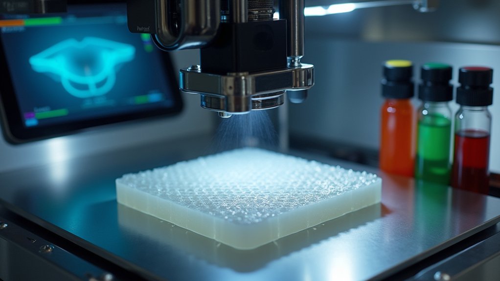
The success of your bioprinted cartilage repair depends on achieving mechanical properties that closely mirror native tissue characteristics.
You’ll need to target an aggregate modulus of approximately 1.8 MPa for articular cartilage and 100-150 kPa for meniscal tissue. Incorporate collagen type II and proteoglycans into your bioinks to replicate the natural extracellular matrix structure.
Design your scaffolds with varying stiffness to reflect zonal differences—circumferential tensile modulus should reach several MPa, while radial direction remains lower.
Add growth factors like TGF-β1 and BMP2 to promote chondrogenic differentiation and enhance mechanical strength.
Test and optimize your bioinks’ rheological properties during printing to guarantee sufficient structural integrity throughout the fabrication process.
Implement Bionic Strategies Based on Natural Cartilage Architecture
By studying cartilage’s three-layer organization, you’ll create bioprinted scaffolds that replicate the superficial zone’s aligned collagen fibers, the middle zone’s random fiber orientation, and the deep zone’s perpendicular arrangement.
These bionic strategies enable precise replication of cartilage architecture, ensuring your scaffolds match native tissue’s functional properties.
You’ll enhance scaffold integration by incorporating advanced bioinks containing collagen types II and aggrecan, which optimize cell behavior and promote superior tissue regeneration.
The biochemical composition of these bioinks directly influences how cells interact with your scaffolds.
Research demonstrates that bionic designs using materials like polycaprolactone successfully replicate complex structures for meniscus regeneration.
Monitor Scaffold Degradation Rates for Optimal Integration
Since scaffold degradation directly impacts tissue integration success, you’ll need to precisely synchronize degradation rates with new cartilage formation to prevent construct failure.
Your bioprinted scaffolds must degrade at controlled speeds that enhance cell behavior while supporting tissue formation throughout the repair process.
Controlled scaffold degradation rates must precisely match tissue formation speed to optimize cell behavior and ensure successful cartilage repair outcomes.
You can achieve ideal integration by selecting materials like polycaprolactone (PCL) and gelatin methacryloyl (GelMA), which offer tunable degradation profiles.
Monitor your scaffolds through in vitro testing to track:
- Mass loss patterns revealing degradation kinetics over time
- Mechanical properties changes showing structural integrity decline
- Cell proliferation responses indicating biological compatibility
Faster degradation rates often stimulate increased cell proliferation and matrix production for cartilage applications.
However, you must balance speed with structural support requirements to guarantee successful tissue integration.
Apply Multi-Zone Printing for Histological Accuracy
While monitoring degradation rates guarantees proper scaffold breakdown, achieving true cartilage repair requires replicating the complex layered architecture of natural cartilage tissue.
You’ll need to implement multi-zone printing techniques that create distinct zones mimicking cartilage’s superficial, middle, deep, and calcified layers. Each zone should contain specific bioink concentrations and growth factors that promote targeted chondrogenic differentiation and extracellular matrix (ECM) production.
Your gradient printing approach must vary mechanical properties across zones to match native cartilage tissue characteristics. This strategy enhances glycosaminoglycan (GAG) production while optimizing cellular behavior in each layer.
Multi-zone scaffolds greatly improve host tissue integration compared to uniform structures, reducing wear and degradation risks. You’ll achieve superior functional outcomes by precisely controlling bioactive substance distribution throughout each zone.
Evaluate Long-Term Cell Viability and Matrix Production
You’ll need to continuously monitor your bioprinted cartilage’s chondrocyte metabolic activity using viability assays to guarantee cells remain functional throughout the repair process.
Track collagen synthesis rates through immunohistochemistry and biochemical assays, as consistent type II collagen production indicates successful chondrogenic differentiation.
Monitor proteoglycan accumulation patterns using Alcian blue and Safranin-O staining to confirm your construct’s developing the proper cartilage matrix composition over time.
Monitor Chondrocyte Metabolic Activity
Although bioprinted cartilage constructs show initial promise, their long-term success depends heavily on maintaining robust chondrocyte metabolic activity throughout the healing process.
You’ll need to regularly assess cell viability using LDH and ATP measurements to track your chondrocytes’ health within the cartilage matrix.
Monitor the expression of chondrogenic markers through these essential techniques:
- Histological staining with Alcian blue and Safranin-O to visualize extracellular matrix production
- Real-time quantitative PCR analysis to measure aggrecan and collagen type II gene expression levels
- Regular metabolic activity assessments to track GAG synthesis and overall cellular function
You should focus on quantifying chondrogenic gene expression using qPCR, as this provides detailed insights into your cartilage constructs’ functionality over time and confirms successful differentiation protocols.
Assess Collagen Synthesis Rates
Tracking collagen synthesis rates provides the most reliable indicator of your bioprinted cartilage’s regenerative potential.
You’ll need to monitor type II collagen production specifically, as it’s the primary component of hyaline cartilage in your bioprinted constructs. Focus on measuring glycosaminoglycan levels, which directly correlate with collagen synthesis and indicate superior tissue quality.
Conduct regular histological analysis to confirm collagen type II expression throughout your construct’s development. This assessment reveals the maturation progress of your engineered cartilage tissue.
Additionally, evaluate the mechanical properties of your bioprinted constructs by testing compressive and tensile strength. These measurements provide vital insights into matrix production effectiveness while ensuring long-term cell viability remains ideal for sustained cartilage regeneration success.
Track Proteoglycan Accumulation Patterns
Because proteoglycans serve as the foundation for cartilage’s compressive strength and water retention capabilities, you must establish systematic monitoring protocols to track their accumulation patterns throughout your bioprinted construct’s development.
Proteoglycan accumulation directly correlates with cell viability and functional cartilage matrix formation in your bioprinted constructs.
Implement these visualization strategies to assess cartilage regeneration:
- Alcian blue staining – Creates vivid blue coloration highlighting glycosaminoglycan distribution patterns across tissue sections
- Safranin-O staining – Produces distinctive red-orange contrast showing proteoglycan density gradients within matrix structures
- Advanced imaging methods – Generates detailed spatial maps revealing three-dimensional proteoglycan deposition architectures
Monitor cell proliferation rates alongside histological staining techniques to optimize your bioink formulations.
This integrated approach guarantees you’re tracking both cellular health and matrix production simultaneously, enabling precise adjustments to growth factor concentrations for enhanced cartilage regeneration outcomes.
Frequently Asked Questions
How Can I Speed up Cartilage Repair?
You can accelerate cartilage repair by incorporating growth factors like TGFβ1 and BMP2 into your bioinks, optimizing scaffold properties through precise 3D printing, and using co-culture strategies with iPSCs.
What Stimulates Cartilage Repair?
Growth factors like TGF-β1 and BMP2 stimulate your cartilage repair by promoting chondrocyte differentiation. You’ll also benefit from mechanical loading, which encourages cellular activity, and co-culturing stem cells to boost glycosaminoglycan production.
What Is the Success Rate of Cartilage Regeneration?
You’ll find current cartilage repair methods have disappointing success rates, with microfracture and autologous chondrocyte implantation failing in 25-50% of cases within ten years, highlighting why you need better regenerative approaches.
How to Reshape Cartilage Naturally?
You can’t actually reshape cartilage naturally once it’s fully formed. However, you can support cartilage health through proper nutrition, targeted exercises, maintaining healthy weight, and avoiding repetitive stress that damages existing cartilage structure.


Leave a Reply