You’ll discover seven cutting-edge vascular network printing techniques revolutionizing tissue engineering: Co-SWIFT method for complex vascular networks, double-crosslinked gelatin-alginate hydrogel printing with enhanced stability, support bath-based bioprinting using smart bioinks, microextrusion coaxial nozzle systems creating core-shell structures, laser-assisted formation achieving 100-micrometer channels, Schiff-base crosslinking for thermal stability, and multi-material core-shell architecture printing. These breakthrough methods enable precise control over vessel geometry, maintain structural integrity at body temperature, and support cell viability for organ transplantation applications that’ll transform regenerative medicine.
Co-SWIFT (Coaxial Sacrificial Writing in Functional Tissue) Method
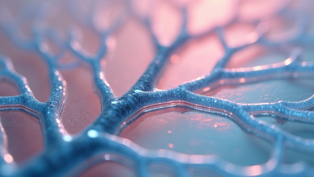
While traditional 3D bioprinting struggles to replicate the complex architecture of natural blood vessels, the Co-SWIFT method developed by Harvard’s Wyss Institute breaks new ground by using coaxial printing to construct sophisticated vascular networks.
You’ll find this technique creates an outer shell of smooth muscle cells with an inner core for fluid flow, giving you precise control over living cells and bioinks arrangement.
The method embeds vascular structures within a transparent hydrogel matrix, letting you test constructs before using collagen for realistic tissue models.
This innovative embedding approach enables thorough testing in transparent matrices before advancing to collagen-based models for authentic tissue replication.
You can create cardiac organ building blocks from human heart cells that beat synchronously after perfusion.
Future applications will enhance capillary networks replication, improving lab-grown tissue functionality for organ transplantation and repair applications.
Double-Crosslinked Gelatin-Alginate Hydrogel Printing
Although traditional gelatin hydrogels lose their structural integrity above 25°C, double-crosslinked gelatin-alginate hydrogels overcome this critical limitation through strategic incorporation of oxidized pullulan and calcium chloride.
You’ll find these enhanced hydrogels deliver superior mechanical properties and thermal stability essential for successful bioprinting applications.
The double-crosslinked system greatly improves biocompatibility while promoting effective cell proliferation and migration throughout your printed constructs. You can create robust vascularized structures that maintain their form during tissue engineering processes.
These hydrogels form interconnected porous structures that facilitate ideal transport of nutrients, oxygen, and metabolites.
When you’re bioprinting complex tissue constructs, this dual crosslinking approach guarantees your gelatin-alginate matrix supports blood vessel integration while maintaining the structural integrity necessary for functional tissue development and long-term viability.
Support Bath-Based Bioprinting With Smart Bioinks
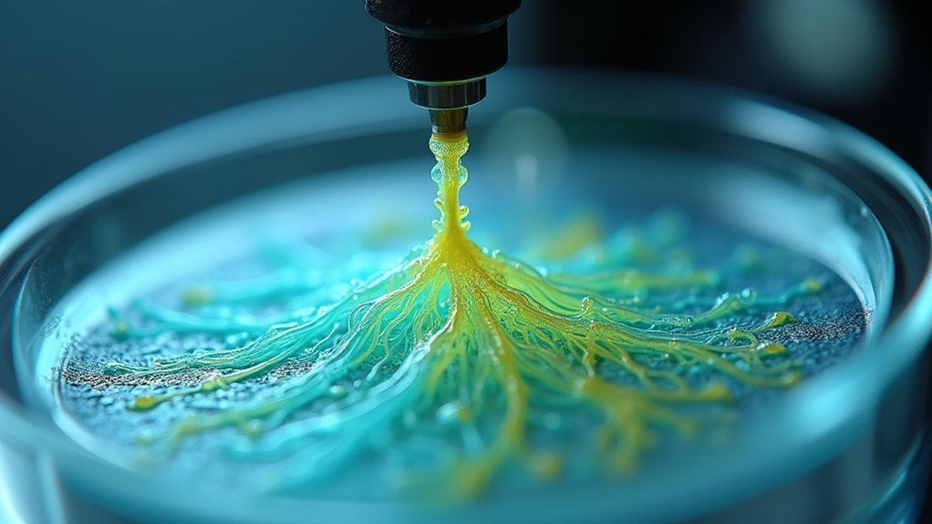
When you’re implementing support bath-based bioprinting with smart bioinks, you’ll need to carefully evaluate bath material properties like viscosity, shear-thinning behavior, and biocompatibility to guarantee ideal printing resolution and cell viability.
Your material selection directly impacts how well the bath maintains structural integrity during complex vascular network formation while allowing smooth bioink extrusion.
You’ll also want to establish robust crosslinking mechanisms that provide immediate gelation upon deposition while maintaining long-term stability throughout the printing process.
Bath Material Selection Criteria
Since smart bioinks require specific environmental conditions to maintain cellular integrity during vascular network printing, you’ll need to carefully evaluate bath material based on several critical criteria.
Your support bath selection must prioritize biocompatible materials that enhance cell viability throughout the bioprinting process.
Consider these essential factors:
- Mechanical properties – The bath material needs ideal viscosity and shear-thinning capabilities to support printed vascular structures while enabling clean removal afterward.
- Thermal stability – Your chosen material must withstand printing conditions without degrading or compromising smart bioinks.
- Biocompatibility – Materials like gelatin and alginate create environments that promote cell growth and vascular integration.
- Degradation rate – The material’s breakdown timeline should align with your vascular structures’ maturation process for successful tissue engineering applications.
Crosslinking Mechanisms For Stability
Once you’ve selected appropriate bath materials that support cellular integrity, implementing effective crosslinking mechanisms becomes your next priority for creating stable vascular networks.
You’ll need crosslinking agents like glutaraldehyde or oxidized pullulan to enhance your bioinks’ mechanical properties. These agents form covalent or ionic bonds that reinforce hydrogel structures, ensuring structural stability during bioprinting processes.
Consider using double-crosslinked hydrogels through sequential crosslinking methods. They’ll provide superior resistance to degradation compared to single-crosslinked systems, making them ideal for tissue engineering applications.
You can also incorporate smart bioinks that respond to external stimuli, creating dynamic vascular constructs that adapt to physiological conditions. This approach allows your vascular networks to maintain functional integrity while supporting complex geometries that mimic natural blood vessel architecture.
Microextrusion Coaxial Nozzle Systems
Microextrusion coaxial nozzle systems represent a breakthrough in vascular network printing, enabling you to simultaneously deposit multiple bioinks through concentrically arranged nozzles.
These systems create core-shell structures that precisely mimic natural vascular architecture while maintaining exceptional cell viability throughout the printing process.
You’ll benefit from four key advantages:
- Dual-material deposition – Print distinct cell types in separate hydrogel layers simultaneously
- Flow rate precision – Control inner and outer material flows for ideal core-shell formation
- Enhanced mechanical properties – Integrate various hydrogel compositions for superior structural integrity
- Geometric flexibility – Fabricate tailored vascular networks matching physiological conditions
The protective hydrogel shell you create encapsulates living cells, promoting functionality during printing.
The protective hydrogel shell encapsulates living cells, maintaining their viability and functionality throughout the entire bioprinting process.
This adaptability makes coaxial nozzle systems essential for advanced tissue engineering applications requiring complex vascular structures.
Laser-Assisted Vascular Structure Formation
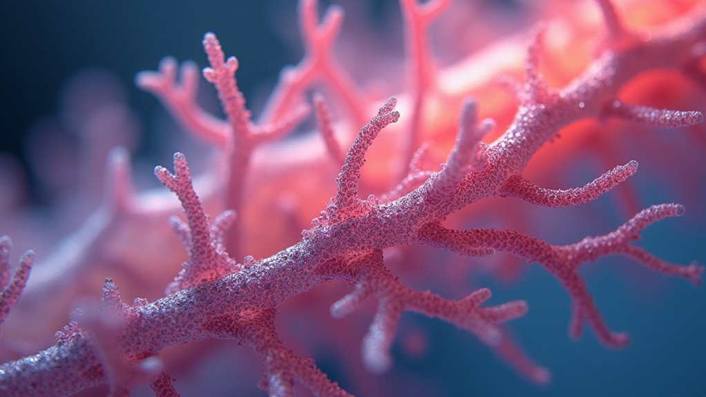
While microextrusion systems excel at creating core-shell structures, laser-assisted bioprinting takes vascular network fabrication to the next level by using focused laser beams to precisely pattern cells and biomaterials with exceptional resolution.
You’ll achieve minimal cellular damage while building complex 3D vascular networks that closely mimic natural blood vessel architecture through layer-by-layer fabrication.
You can control laser intensity and duration to enhance your hydrogel’s mechanical strength and stability.
Laser-assisted printing enables you to incorporate multiple cell types simultaneously, creating heterogeneous structures that replicate native tissue composition.
You’ll produce perfusable channels as small as 100 micrometers in diameter, essential for maintaining tissue viability in engineered organs.
This technique delivers the precision needed for functional vascular structures.
Schiff-Base Crosslinking for Enhanced Stability
You can enhance your bioprinted vascular networks’ structural integrity by implementing Schiff-base crosslinking with oxidized pullulan, which forms stable imine bonds between aldehyde and amine groups in your hydrogel matrix.
This crosslinking mechanism doesn’t just improve mechanical properties—it creates a thermally stable network that maintains its shape and function under physiological conditions.
You’ll find that oxidized pullulan’s aldehyde groups react efficiently with gelatin’s amino groups, producing crosslinked structures that resist thermal degradation while preserving biocompatibility.
Oxidized Pullulan Mechanisms
The formation of Schiff-base bonds between oxidized pullulan and gelatin creates a robust double-crosslinking network that dramatically enhances hydrogel stability for vascular printing applications.
You’ll achieve superior mechanical properties when oxidized pullulan’s aldehyde groups react with gelatin’s amino groups through Schiff-base reactions.
This crosslinking mechanism delivers four key advantages for vascular tissue engineering:
- Enhanced structural integrity – Your bioprinted vascular structures resist degradation in physiological conditions
- Tunable gelation properties – You can customize mechanical characteristics to mimic natural extracellular matrix
- Maintained biocompatibility – Cell attachment and proliferation aren’t compromised during crosslinking
- Extended functional lifespan – Your hydrogels maintain performance longer than traditional formulations
You’ll find this double-crosslinking approach essential for creating durable, functional vascular networks that support tissue regeneration.
Thermal Stability Enhancement
Beyond mechanical improvements, Schiff-base crosslinking dramatically boosts thermal stability in your bioprinted vascular constructs. This crosslinking method transforms standard gelatin-alginate hydrogels into robust matrices that maintain structural integrity at physiological temperatures. You’ll achieve superior mechanical properties while preserving essential biocompatibility for tissue engineering applications.
| Property | Standard Hydrogels | Schiff-Base Crosslinked |
|---|---|---|
| Thermal Stability | Poor at 37°C | Excellent at 37°C+ |
| Structural Integrity | Degrades quickly | Maintains form |
| Cell Interaction | Limited | Enhanced |
| Mechanical Strength | Weak | Notably improved |
| 3D Bioprinting Success | Challenging | Reliable |
Your vascular networks benefit from enhanced cell interaction within the oxidized pullulan matrix. This crosslinking approach enables successful 3D bioprinting of complex geometries that withstand physiological conditions, making your constructs viable for practical physiological applications in regenerative medicine.
Multi-Material Core-Shell Architecture Printing
While traditional 3D printing methods struggle to replicate the complex layered structure of natural blood vessels, multi-material core-shell architecture printing revolutionizes vascular network creation by simultaneously depositing distinct materials through coaxial nozzle systems.
This printing technique creates vascular structures with smooth muscle cell shells surrounding cores optimized for fluid flow. You’ll control vessel diameters by adjusting printing speeds and ink flow rates through independently controllable channels.
The process offers four key advantages:
- Enhanced cell integration – You can infuse endothelial cells into cores after removing initial materials.
- Improved nutrient transport – Core-shell designs optimize oxygen and nutrient delivery.
- Customizable architecture – You’ll create patient-specific vascularized organ models.
- Superior functionality – Maintains cell viability while producing functional constructs.
Frequently Asked Questions
Which Bioprinting Technology Is Most Used to Create Bioprinted Vascular Networks?
You’ll find extrusion-based bioprinting is the most widely used technique for creating bioprinted vascular networks. It’s popular because you can easily fabricate complex structures quickly and cost-effectively compared to other methods.
What Is 4D Printing for Artificial Blood Vessels?
You’ll use materials that change shape over time when exposed to stimuli like temperature or moisture, creating blood vessels that can self-assemble and adapt after printing, mimicking natural vascular remodeling processes.
Can You 3D Print Veins?
You can 3D print veins using bioprinting techniques like coaxial printing. You’ll create layers with endothelial and smooth muscle cells using bioinks containing hydrogels. These printed structures can function and respond to cardiac drugs effectively.
What Are the Techniques of Bioprinting?
You’ll find bioprinting uses several techniques: extrusion-based printing for speed and cost-effectiveness, inkjet-based for precision, laser-assisted methods, coaxial printing for cell encapsulation, and embedded printing within hydrogel matrices.
Leave a Reply