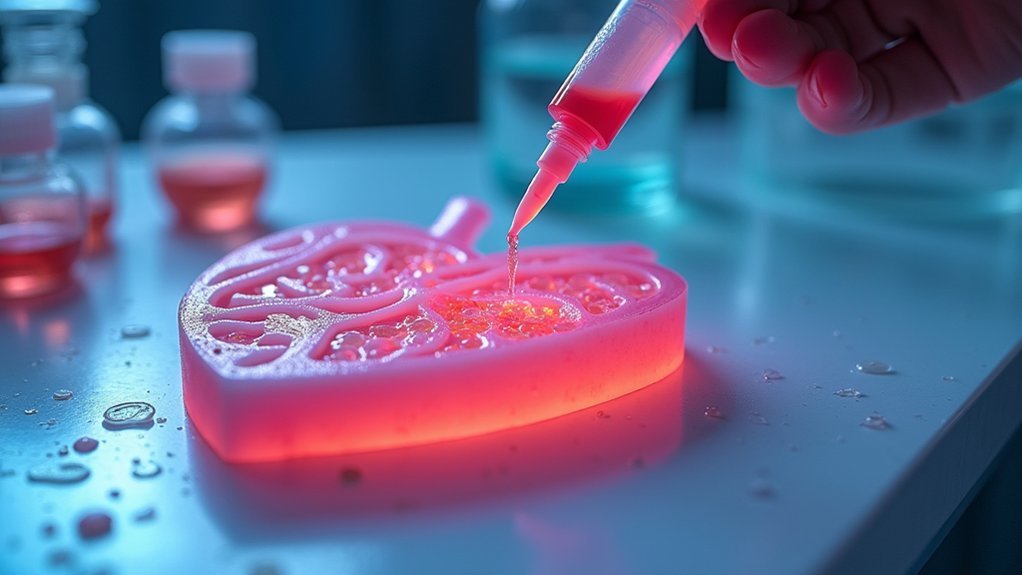
You’ll need bioinks combining natural ECM components like collagen and decellularized extracellular matrix with synthetic polymers to achieve the critical 8-17 kPa stiffness range that cardiomyocytes require. Your formulation should include conductive materials like carbon nanotubes or graphene to reach 0.1-10 S/m conductivity for proper electrical signaling. The bioink must maintain 100-500 mPa·s viscosity during printing while incorporating growth factors and biochemical cues for cell differentiation. These hybrid systems require specific cross-linking mechanisms and rheological properties to create functional cardiac constructs.
Understanding the Cardiac Microenvironment for Bioink Design
When you’re designing bioinks for cardiac tissue engineering, you must first understand the complex cellular architecture that defines the heart’s microenvironment.
The cardiac microenvironment contains cardiomyocytes and cardiac fibroblasts that communicate through intricate biochemical cues and mechanical signals. Your bioink formulation needs to replicate the extracellular matrix properties, particularly achieving ideal matrix stiffness around 10 kPa for proper cardiomyocyte maturation.
You’ll need to incorporate growth factors and signaling pathways like Wnt and Notch to guide cell differentiation effectively.
The inclusion of decellularized extracellular matrix enhances cellular behavior by mimicking native tissue conditions.
Additionally, you must consider how electrical stimulation promotes cell-cell coupling and improves contractility in your printed constructs, ensuring the bioink supports proper ion channel development.
Natural Biomaterial Components in Cardiac Bioink Formulations
When you’re formulating cardiac bioinks, you’ll find that collagen serves as the backbone material due to its natural abundance in heart tissue and its ability to provide structural integrity while promoting cell attachment.
You can enhance these collagen-based formulations by incorporating decellularized extracellular matrix components that retain the native biochemical signals essential for cardiomyocyte function and differentiation.
These ECM-derived elements work synergistically with collagen to create a more authentic cardiac microenvironment that supports proper tissue development and maturation.
Collagen-Based Matrix Properties
As the backbone of cardiac tissue architecture, collagen forms the foundation for many successful bioink formulations designed for heart tissue engineering applications.
When you’re developing cardiac bioinks, you’ll find that Type I collagen enhances mechanical properties by achieving ideal stiffness levels between 8-17 kPa, which directly supports cardiomyocyte maturation. You can customize collagen-based hydrogels through physical or chemical crosslinking methods, allowing you to control degradation rates and replicate native cardiac environments.
Collagen’s role extends beyond structural support—it facilitates cardiomyocyte alignment, ensuring proper electrical conductivity and coordinated contractions in engineered heart tissues.
The extracellular matrix (ECM) component’s exceptional biocompatibility promotes cell adhesion, proliferation, and differentiation while supporting vascularization, making it essential for integrating functional bioprinted cardiac constructs.
ECM-Derived Bioink Components
Beyond collagen’s foundational role, you’ll discover that incorporating diverse ECM-derived components creates bioink formulations with enhanced bioactivity and structural complexity for cardiac applications.
These natural biomaterials work synergistically to replicate your target tissue’s native environment.
Key ECM-derived bioinks components include:
- Fibrin networks – Crosslinked fibrinogen forms fibrous hydrogels that promote cellular interactions and enhance vascularization within your engineered constructs.
- Gelatin methacryloyl (GelMA) – Offers excellent cytocompatibility with tunable mechanical properties that match native myocardium stiffness.
- Decellularized ECM (dECM) – Retains tissue-specific biochemical signals that enhance cell adhesion and drive differentiation toward cardiac phenotypes.
You’ll find that combining these components allows precise control over your bioink’s mechanical properties while maintaining the bioactive cues essential for successful cardiac tissue engineering.
Synthetic Polymers for Enhanced Printability and Function
When you’re formulating cardiac bioinks, synthetic polymers offer precise control over mechanical properties that you can’t achieve with natural materials alone.
You’ll find that these engineered components enhance printability through improved rheological behavior while maintaining the biocompatibility essential for cardiac cell survival and function.
Your choice of cross-linking strategy becomes critical, as it determines both the structural integrity of printed constructs and the degradation timeline that supports tissue maturation.
Mechanical Property Control
While natural biomaterials offer excellent biocompatibility, synthetic polymers provide unprecedented control over mechanical properties that’s essential for cardiac bioprinting success. You can precisely tune your bioinks using polyethylene glycol and poly(lactic acid) to match native cardiac tissue stiffness of 8–17 kPa, directly supporting cardiomyocyte function.
Methacrylated polymers like GelMA enhance cytocompatibility while maintaining excellent printability characteristics. Your strategic polymer ratio adjustments enable ideal viscosity control, preserving cell viability during extrusion.
You’ll achieve superior mechanical support by engineering degradation rates that align with natural tissue remodeling processes.
- Stiffness matching: Your bioprinted scaffold mirrors the exact elasticity of beating heart muscle
- Cellular harmony: Cardiomyocytes contract rhythmically within perfectly tuned mechanical environments
- Temporal precision: Scaffold dissolution synchronizes seamlessly with new tissue formation
Biocompatibility and Degradation
Although synthetic polymers like PEG and PLA might seem less biologically intuitive than natural materials, they’ve revolutionized cardiac bioprinting through their exceptional biocompatibility when properly engineered.
You can enhance these bioink formulations by incorporating bioactive components that promote cell adhesion and proliferation within your engineered cardiac tissues.
The key advantage lies in controlling degradation rates to match tissue regeneration timelines, ensuring your scaffolds support cellular growth throughout the healing process.
As synthetic polymers gradually break down, cells produce their own extracellular matrix (ECM), creating seamless tissue integration.
This controlled biodegradability allows you to maintain ideal mechanical properties while facilitating the natural replacement of synthetic materials with native tissue components, ultimately achieving superior biocompatibility in your printed cardiac constructs.
Cross-linking Enhancement Strategies
Since achieving ideal printability requires precise control over material properties, cross-linking enhancement strategies represent the cornerstone of successful synthetic polymer bioink formulations for cardiac applications.
You’ll find that chemical modifications dramatically improve your bioink properties. Methacrylation transforms gelatin into GelMA hydrogels with tunable mechanical properties that mimic myocardial stiffness. Incorporating photoinitiators in your synthetic polymers enables rapid UV cross-linking for creating intricate 3D cardiac structures with exceptional resolution.
Key cross-linking enhancement approaches include:
- Chemical modification – PEG and PLA polymers gain superior cross-linking capabilities through functional group additions.
- Hybrid formulations – Combining synthetic polymers with natural biomaterials balances mechanical strength and cell attachment.
- Ratio optimization – Fine-tuning polymer concentrations achieves low printing viscosity while ensuring adequate post-cross-linking strength for functional cardiac tissue.
Hybrid Bioink Systems Combining Natural and Synthetic Materials
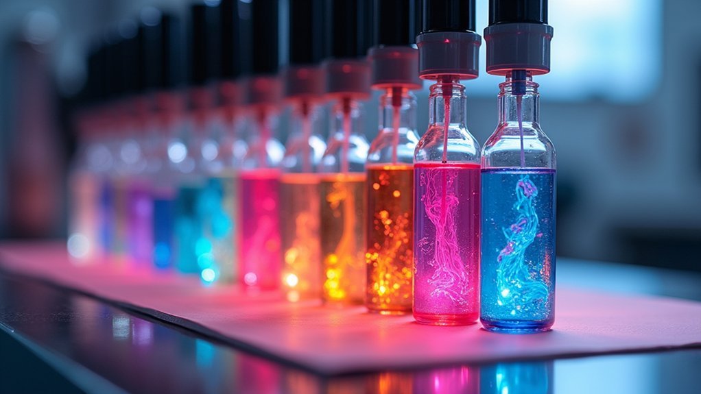
When developing bioinks for cardiac tissue engineering, you’ll find that hybrid systems combining natural and synthetic materials offer the most promising approach to achieving the complex requirements needed for functional heart tissue. These formulations strategically combine gelatin and collagen with synthetic polymers like PEG to enhance mechanical properties and biocompatibility.
| Component Type | Key Benefits |
|---|---|
| Natural Materials | Enhanced cell attachment and biocompatibility |
| Synthetic Polymers | Tunable mechanical properties and printability |
| dECM Integration | Improved cardiomyocyte differentiation signals |
You can tailor these hybrid bioink systems to match heart tissue’s 8-17 kPa stiffness range, promoting proper cell function. The tunable viscosity guarantees ideal printability while maintaining cell viability during 3D bioprinting. Adding decellularized extracellular matrix provides vital biochemical cues that enhance cardiomyocyte differentiation and tissue maturation.
Cell Integration Strategies in Cardiac Bioinks
The success of hybrid bioink formulations ultimately depends on how effectively you integrate living cells within these materials to create functional cardiac tissue.
Effective cell integration within hybrid bioink formulations is the critical determinant of functional cardiac tissue engineering success.
Your cell integration strategies in cardiac bioinks must account for multiple factors that influence tissue formation. When developing your approach, you’ll need to create ideal conditions for cardiomyocytes and cardiac fibroblasts to thrive.
Incorporating decellularized extracellular matrix enhances bioactivity while providing structural support. You can also embed biochemical cues to guide stem cell differentiation toward cardiac lineages.
Consider these essential integration strategies:
- Tune mechanical properties to approximately 10 kPa stiffness for ideal cardiomyocyte maturation
- Design multi-material bioinks that spatially distribute different cell types throughout the construct
- Encapsulate growth factors within hydrogel networks to sustain cellular development over time
Rheological Properties Essential for Cardiac Tissue Printing
Although cellular viability remains paramount in cardiac bioprinting, you must carefully balance rheological properties to achieve both printability and biological function.
Your bioink requires specific viscosity characteristics—maintaining 100-500 mPa·s before printing for precise deposition while demonstrating yield stress between 10-100 Pa for shape fidelity. You’ll need shear-thinning behavior that allows smooth extrusion under stress while recovering viscosity post-deposition.
The mechanical stiffness of your hydrogel formulation should mimic native cardiac extracellular matrix properties, with storage and loss moduli around 8-17 kPa to promote ideal cardiomyocyte maturation.
Incorporating materials like GelMA enhances printability through controlled shear-thinning responses. You must guarantee these rheological properties support both structural integrity during printing and proper cardiac tissue development afterward.
Cross-linking Mechanisms for Structural Integrity
Once your bioink achieves ideal rheological properties, you’ll need robust cross-linking mechanisms to lock these characteristics into place and provide lasting structural integrity.
Robust cross-linking mechanisms are essential to transform printable bioinks into structurally stable cardiac tissue constructs with lasting integrity.
Cross-linking mechanisms transform your bioink formulations from printable materials into stable cardiac tissue constructs with precise mechanical properties.
You can achieve cross-linking through several approaches:
- Photopolymerization – Expose GelMA bioinks to UV or visible light for rapid, controlled solidification
- Ionic gelation – Trigger calcium-mediated cross-linking for gentle, cell-friendly stabilization
- Thermal processing – Apply controlled temperature changes to activate protein-based cross-linking
Target around 10 kPa stiffness to mimic native myocardial extracellular matrix, optimizing cardiomyocyte maturation.
Proper cross-linking enhances bioactivity, promoting essential cell adhesion, proliferation, and differentiation for functional cardiac constructs.
Electrical Conductivity Enhancement in Cardiac Bioinks
Beyond structural stability, your cardiac bioinks must conduct electrical signals effectively to enable the synchronized contractions that define functional heart tissue.
You’ll need to incorporate conductive materials like carbon nanotubes, graphene, or polyaniline into your formulations to achieve ideal electrical conductivity. Research shows bioinks with conductivity ranging from 0.1 to 10 S/m greatly improve bioprinted cardiac constructs by enhancing cell-to-cell communication.
Consider using conductive hydrogels such as gelatin methacryloyl modified with conductive nanoparticles, which maintain essential bioactivity while delivering enhanced electrical properties.
This approach promotes better maturation of stem cell-derived cardiomyocytes, resulting in increased contractile force and more physiological responses in your engineered heart tissues.
Mechanical Property Optimization for Heart Tissue Mimicry
While electrical conductivity enables proper signal propagation, your bioinks must also replicate the unique mechanical environment of native heart tissue to achieve functional cardiac constructs.
You’ll need to match the 8-17 kPa stiffness range of myocardial tissue to promote suitable cardiomyocyte maturation and functionality.
Your bioink formulation requires careful balance of several key mechanical properties:
- Low viscosity during extrusion – ensuring smooth printing flow while maintaining structural integrity for complex cardiac geometries
- Suitable porosity levels – creating interconnected networks that allow nutrient diffusion and oxygen transport to embedded cardiomyocytes
- Tunable biodegradability – programming gradual degradation as cells produce their own extracellular matrix
Chemical modifications like gelatin methacrylation can enhance your bioink’s mechanical properties while promoting cell attachment, creating the ideal environment for cardiac tissue development.
Frequently Asked Questions
What Is the Bioink Used in Bioprinting?
You’ll typically use hydrogel-based bioinks containing natural biomaterials like collagen, gelatin, or fibrin. These materials provide cell-friendly environments with appropriate viscosity for printing while maintaining viability during the fabrication process.
Can You Bioprint a Heart?
You can’t bioprint a complete, fully functional heart yet. Current technology lets you create cardiac tissue constructs and small heart patches, but scientists haven’t achieved the complexity needed for whole hearts.
Can Mucus Based Bioink Be Used to Print and Grow Lung Tissue?
You can use mucus-based bioinks to print lung tissue successfully. They’ll mimic natural lung ECM, enhance cell adhesion, support airway formation, and promote ciliary differentiation while maintaining excellent printability for complex structures.
Which Bioprinting Technology Is Most Used to Create Bioprinted Vascular Networks?
You’ll find inkjet-based bioprinting is most commonly used for creating vascular networks because it precisely deposits bioinks in droplets, achieving high cell viability and excellent construct resolution for intricate vascular structures.

Leave a Reply